Chapter: Wounds and ulcerations
Article: 2 of 7
Update: Mar 10, 2021
Author(s): Ott, Hagen
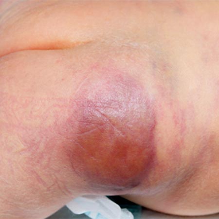
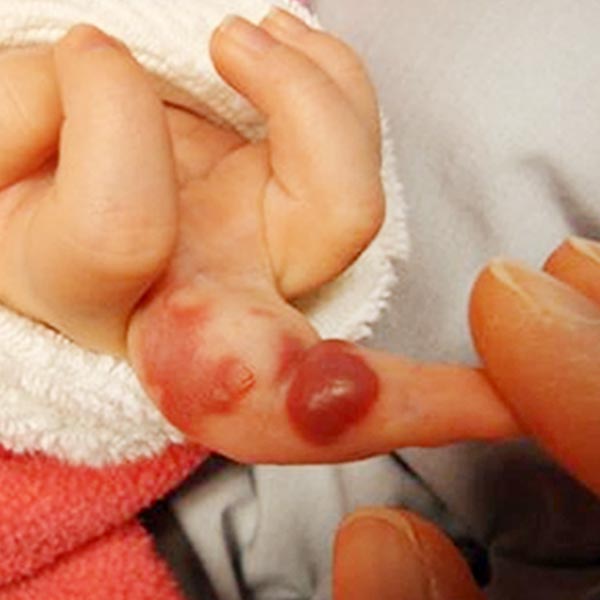
Skin defects and ulcerations are among the typical, although not very common, complications of vascular anomalies. They are observed in the natural history of patients with infantile hemangiomas (IH). Thus, infantile hemangiomas evaluated in specialized consultations have ulcerations in approximately 15% to 25%.
These often develop towards the end of the first, rapid growth phase (proliferation phase) between the fourth and eighth month of life. They are mainly observed in body folds (e.g., the genito-anal area), on the lip and in the head and neck region. An increased risk of ulceration is also found in the case of segmental infantile hemangiomas and large infantile hemangiomas with superficial and deep portions. Interestingly, later ulcerating infantile hemangiomas often show whitish discoloration at the rim of the lesion and/or crusting in the central portion in the early phase.
Depending on vessel architecture and location, arteriovenous malformations may also develop ulcers and gangrenous changes near the nidus in their spontaneous course or after surgical therapy. This is particularly common during the spontaneous course on the lower extremities and here especially on the lower leg and foot because of the additional high hydrostatic pressure there.
Extensive vascular malformations, especially lymphatic malformations, can lead to irritation and inflammation of affected areas in the region of newly formed skin folds. This is called intertriginous dermatitis and is accompanied by softening (maceration) and superficial skin defects (erosions). As a consequence, the risk of secondary infections with bacteria and/or fungi is significantly increased.