Chapter: Lymphatic malformations
Article: 3 of 14
Update: Mar 15, 2021
Author(s): Meyer, Lutz
The lymphatic system, like all structures of the body, develops in a complex process during embryonic development. During this process, various intermediate stages are passed through, controlled by diverse factors, until the mature system of lymphatic vessels is formed, which returns body fluids and cells that have left the vascular system back into the venous system.
Knowledge of the various stages of development makes it understandable why a circumscribed microcystic or macrocystic lymphatic malformation (hereafter “LM”) develops preferentially in certain areas of the body. A lymphatic malformation is never intracerebral, subglottic, intratracheal or intraarticular, very rarely intraosseous (as in Gorham-Stout syndrome) but occurs all the more frequently in the region of the facial skull and neck, sometimes extending into the mediastinum or from retroperitoneally to the soft tissues of the pelvis and thigh. However, lymphatic malformation can also affect just the skin or the skin and subcutis. This is sometimes in very circumscribed areas measuring only a few centimeters (the term lymphangioma circumscriptum is used clinically for this, although it is not in the ISSVA classification).
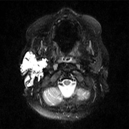
A classic lymphatic malformation consists of a circumscribed lesion with cystic dilations of lymphatic vessels of varying size, a thin wall with many septations, and is filled with lymph fluid without drainage into the normal lymphatic system. Lymph fluid (clear yellow or milky white in the case of chyle) forms and collects in the cysts, causing the cysts to grow to different sizes. When the cyst size exceeds 2 cm (in some definitions 1 cm), it is referred to as a macrocystic LM; smaller cysts are referred to as microcystic LMs. In many cases cysts of both groups are present (then called “mixed cystic LM” according to the ISSVA classification). Small feeding blood vessels usually run in the cyst walls; these may bleed during surgical procedures and occasionally cause the LM to be misclassified as a mixed vascular malformation (e.g., lymphatic-venous malformation LVM). Nevertheless, a true LVM may also be present.
A rare and particularly extensive form of LM is generalized lymphatic anomaly (GLA), which was formerly called lymphangiomatosis. In this case, bone, intestine and parenchymal abdominal organs as well as the lungs may be involved.
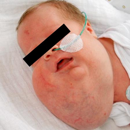
A lymphatic malformation displaces the surrounding tissue. This not only causes outward swelling, but relocates surrounding tissue and may also stress bone tissue. A large lymphatic malformation in the face can alter the direction of growth and bone strength of the facial skull, with sometimes severe dysplasia and typical deformities.
A large retroperitoneal lymphatic malformation may affect bowel passage or bladder capacity and emptying.
In the thigh, a lymphatic malformation can develop between muscle groups and cause displacement, usually without symptoms. An LM in the floor of the mouth between and within the numerous small muscles of the tongue often gives the impression of diffuse infiltration and macroglossia.
Cutis and mucous membranes show specific phenomena. An intracutaneous LM, also called lymphangioma circumscriptum, usually located on the extremities, shoulders, or buttocks, may develop scattered or even multiple clusters of small vesicles a few millimeters in diameter.
Initially they are filled with clear lymph and, as they progress, they are increasingly intermingled with blood and then become shimmering red.
Similarly, vesicles filled with clear or bloody lymph may appear on the body of the tongue and sublingually in the mucosa, sometimes combined with further vesicular rashes on the mucosa of the cheek pouches.
Lymphatic malformations of the pharynx and larynx exhibit frogspawn-like collections of usually clear cysts of the mucosa. In the larynx, cysts may extend into the vocal cords but never below the vocal cord level. If the parapharyngeal and retropharyngeal tissues are infiltrated with cysts and frogspawn-like mucosal cysts in the vallecula, in the larynx up to the vocal cords as well as paralaryngeally, dangerous narrowing of the upper airways may occur. Such an obstruction of the airways may also result from compression of the upper airways by large lymphatic cysts in the neck, especially if this expands rapidly due to hemorrhage or bacterial superinfection.
The extent of filling of each lymphatic malformation cyst is not always the same. What factors cause these variations is not yet sufficiently known. However, clinical experience shows that systemic infections, especially of the respiratory or GI tract, can trigger greater filling of cysts. After the infection has subsided, the volume of the individual cysts usually decreases again.