CLOVES syndrome (CLOVES: Congenital Lipomatous Overgrowth, Vascular malformations, Epidermal nevi, Skeletal anomalies) is characterized mainly by the combination of regional overgrowth with a strong soft tissue component (fibrolipomatous overgrowth) and vascular malformations of capillary, venous, lymphatic and also (more rarely) arteriovenous type.
Genetic basis
CLOVES syndrome occurs sporadically (not familial). It is based on a genetic mosaic. It is caused by somatic (present only in affected tissue) autosomal dominant gain-of-function mutations in the oncogene PIK3CA. Mutational constitutive activation of PIK3CA leads to overactivation of the PI3K(phosphatidylinositol 3-kinase)/AKT/mTOR pathway and causes enhanced cell growth and anti-apoptosis.
Clinical presentation
The regional overgrowth with predominant soft tissue component is already present at birth and most often affects the feet. The “CLOVE foot” has a characteristic appearance: widening especially of the forefoot (“ballooned hyperplasia”), fibroadipose soft tissue proliferation, sandal gap.
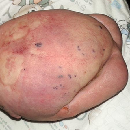
Single or multiple toes or rays may be affected by the overgrowth. In contrast to Proteus syndrome, despite the often massive overgrowth of individual bones, their shape is largely preserved. Furthermore, lipomatoses (circumscribed fat hyperplasias) are found, sometimes over large areas, especially on the trunk. Often patients with CLOVES syndrome also show concomitant lipoatrophies, these mostly affecting the arms. The underlying mechanism of adipose tissue dysregulation is not yet understood.
Epidermal nevi appear as skin-colored to gray-brown raised, striated skin changes with a velvety soft, scaly sometimes papillomatous surface.
Vascular malformations can involve capillaries (CM), veins (VM), and/or lymphatic vessels (LM); furthermore, arteriovenous malformations (AVM) may occur more rarely. Capillary malformations (initially often purplish, later fading) in the trunk area are characteristic. Venous malformation often results in large marginal veins that may extend into the thorax. The arteriovenous malformations are often located near or interspersed in the lipomatous masses; they are also clustered paraspinally.
Therapy
Multidisciplinary and symptomatic
Assessment and treatment (especially of marginal veins) of vascular malformations and, if necessary, thrombosisprophylaxis in risk situations.
Because of the asymmetric growth of the extremities, orthopedic care; aids (corset, shoes), physiotherapeutic measures
Growth control by epiphysiodesis and resection of foot or hand rays if necessary
Individual therapy trials with mTOR inhibitors are described
Complications
Cave: Pulmonary embolism as a result of deep vein thrombosis and thromboembolism