Chapter: Lymphatic malformations
Article: 7 of 14
Update: Mar 15, 2021
Author(s): Meyer, Lutz
The wide spectrum of benign and malignant soft tissue or bone tumors must always be included in the differential diagnosis. If too much focus is placed on vascular malformations, it is possible to lose sight of malignant tumors, for example, and lose valuable time. An important component is simple finger compression of the lesion. A cystic lymphatic malformation (LM) is more likely to be soft or elastic. Tumors (including vascular tumors) feel hard on palpation because they are solid (tightly stacked cells) as opposed to lymphatic malformations (fluid-filled cysts). In contrast, primary lymphedema is soft (initial stage) and less circumscribed.
It is not always easy to differentiate a lymphatic malformation from other variants of vascular malformations such as venous malformations (VM) or arteriovenous malformations (AVM) as well as the group of vascular tumors (different hemangiomas, etc.). Especially in the immediate postnatal period, it is sometimes difficult to make the correct diagnosis in the case of facial swelling because subcutaneous infantile hemangiomas or AVM, for instance, may only develop typical clinical features at a later stage of development. Imaging, such as MRI, is helpful when it is necessary based on symptomatology or for differential diagnosis. Intra-orbital malformations are often particularly difficult to classify if no other symptoms are externally discernible. Even sonography does not always provide an unequivocal diagnosis. Sometimes only intraoperative findings and histology will enable the diagnosis to be made. A congenital intra-orbital meningocele may be confused with lymphatic malformations. If this is opened intraoperatively without being noticed, a cerebrospinal fluid fistula may result, which is not easy to close surgically.
In principle, other cystic processes (e.g., lateral branchiogenic cervical cyst, a residual cervical sinus or median cervical cyst, a residual thyroglossal duct, epidermoid, dermoid cysts, other ciliated cysts) should also be considered, especially in the neck area.
Partially cystic degenerated tumors, in particular, may contain parts reminiscent of a lymphatic malformation (e.g. teratoma, congenital rhabdomyosarcoma, synovial sarcoma, angiomatoid fibrous histiocytoma, epithelioid sarcoma), but they nearly always contain solid portions lacking in lymphatic malformations.
Differential diagnosis may become even more difficult when mixed forms of a lymphatic malformation with, for example, a venous malformation (LVM) occur within a tumorous swelling. There seem to be cases which present postnatally as typical cystic lymphatic malformation on the head and neck and increasingly develop venous malformation components in remaining remnants over the course of years. Existing lymphovenous anastomoses probably play a role in this situation.
Patients with complex syndromes may have a lymphatic malformation as one component among other vascular malformations. Thus, there are numerous possible combinations. In CLOVES syndrome, cystic-lymphatic and venous malformation components often coexist.
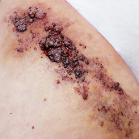
In blue rubber bleb nevus syndrome (Bean syndrome), for example, cystic lymphatic malformations may be present in addition to multiple venous malformations of the skin, visceral organs, or even very rarely of the musculature. The basic diagnostic difference between an LM and a VM is the contrast enhancement on imaging: only the lymph-containing thin wall enhances in an LM, whereas the entire blood-containing lesion enhances in a VM.