Chapter: Venous malformations
Article: 6 of 13
Update: Mar 25, 2021
Author(s): Barbera, Letterio Christian
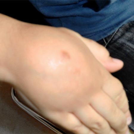
Visible ectatic and bluish vessels, which increase in size on standing, are equally characteristic of varicosis and venous malformation. In both diseases, the affected parts of the body can swell and cause a feeling of heaviness. Nevertheless, history-taking as well as physical and sonographic examination enable the physician to differentiate between the two entities. Distinguishing features (see table) help in making the correct diagnosis, whereby not a single criterion but the overall picture is important.
| Feature | Varicosis | Venous Malformation |
|---|---|---|
| Frequent localization | Leg medial and calf | Leg on its lateral side, usually only one side |
| Patient age | Over 18 years | Starting in childhood |
| Appearance of the vessels | Meandering, tubular , small caliber changes | Plexiform, irregular pattern, large differences in caliber |
| Skin discoloration | Inner ankle, brown | Over the venous malformation bluish discoloration |
| Depth extension | Subcutaneous | Intracutaneous, subcutaneous, subfascial, intramuscular |
| Reflux (Duplex ultrasound) | Can be induced over the entire length of the varicose veins | Often only short segments |
Naturally, a 15-year-old girl could have primary greater saphenous vein varicosis and a 70-year-old man might notice a venous malformation for the first time. Similar examples apply to the other characteristics. However, this is not generally the rule, which is why unusual findings should be critically evaluated, taking into account the entire picture in order to avoid wrong treatments.
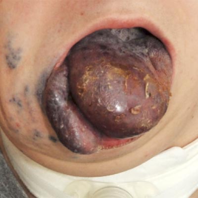
Common features of soft tissue and vascular tumors are increase in size, hypervascularity, growth across tissue layers and symptoms. Especially subfascially located venous malformations are sometimes difficult to distinguish from benign and malignant soft tissue tumors without imaging. Even modern cross-sectional imaging techniques do not allow a sufficiently reliable differentiation to be made between venous malformation and neoplasia in selected cases. Since soft tissue tumors usually grow faster, a second MRI or CT scan after 3–6 months may be crucial. A biopsy is indicated if there are doubts about possible malignancy of the process. Understandably, attention should be paid to obtaining a representative piece of tissue, taking into account the increased risk of bleeding. If other typical features of a congenital vascular malformation are present, such as early onset, long-term medical history, subcutaneous localization, extension over the entire extremity, histologic examination is usually not necessary. Solid tumors with a large number of vessels are, in most cases, easily distinguishable from venous malformations by means of MRI.
Hemangiomas represent a special form of vascular tumors. These may be visible at birth (congenital hemangiomas) or more frequently in the first days of life, they usually grow rapidly, frequently affect the face and generally regress during the first years of life (infantile hemangiomas). Thus, time of first appearance and regression are important distinguishing features in respect of venous malformations. Both sonographically and in MRI, the hollow, blood-filled vascular spaces of venous malformations can be readily distinguished from solid, highly perfused infantile hemangiomas. A congenital hemangioma (NICH, RICH), however, can sometimes be difficult to differentiate from other solid tutors.
Prominent vascular convolutes that grow over the years occur in post-traumatic AV fistulas or dialysis shunts and can be misinterpreted as venous malformations. There are meandering veins with a large diameter and thick walls, which are the result of increased flow and pressure (“fast-flow lesions”).
In contrast to a venous malformation, there is a spontaneous strong blood flow. This is perceived clinically as buzzing (“thrill”) and on duplex sonographically as a holosystolic, high-frequency signal.