Chapter: Imaging
Article: 1 of 10
Update: Feb 05, 2021
Author(s): Müller-Wille, René
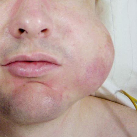
The correct diagnostic classification of a vascular anomaly (ISSVA classification) is essential because therapeutic strategies are differentiated. In addition to medical history and physical examination, radiological imaging plays a vital role in the differential diagnostic work-up of vascular anomalies. The imaging techniques used include sonography, magnetic resonance imaging (MRI), digital subtraction angiography (DSA), phlebography/varicography, conventional X-rays and computed tomography (CT).
| Procedure | Description | Disadvantages |
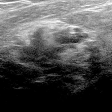
| Sonography |
|
|
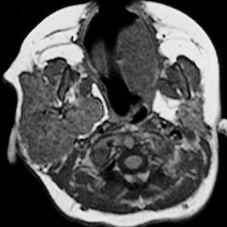
| Magnetic resonance imaging (MRI) |
|
|
| Digital subtraction angiography (DSA) |
|
|
| Phlebography/varicography |
|
|
| Conventional radiography |
|
|
| Computed tomography (CT) |
|
|