Chapter: Imaging
Article: 9 of 10
Update: Feb 05, 2021
Author(s): Wohlgemuth, Walter A.
Intranodal lymphangiography (INL) is used for direct imaging of central conducting abdominal and thoracic lymphatic vessels after injection of an oily X-ray contrast medium (Lipiodol) into inguinal lymph nodes under ultrasound guidance. This new procedure has practically replaced the conventional bipedal lymphangiography, in which a lymph vessel on the back of the foot is surgically prepared and injected with dye.
Opacification of the lymph vessels after sonographic insertion of a thin needle into inguinal lymph nodes on both sides at the border between medulla and cortex of the lymph node must be performed very slowly, preferably using injection pumps, in order to avoid contrast medium leakage from the thin-walled lymph vessels. The outflow of the contrast medium through the central lymphatic ducts of the pelvis, abdomen and chest to the entrance in the left venous angle is anatomically variable. Illustration of the individual anatomy is performed by fluoroscopy.
Indications for intranodal lymphangiography are found in all pathologies of large lymphatic ducts in the body, including central conducting lymphatic anomaly (CCLA) and lymphatic malformations in the abdomen or mediastinum. Postoperative lymphatic leakages are also very easy to visualize and, if necessary, to treat.
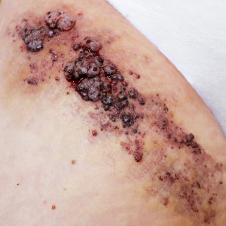
Central conducting lymphatic anomaly (CCLA): This is defined by congenital disorders (stenosis or occlusion) of the central lymphatic ducts, especially the cisterna chyli and the thoracic duct. It can be accompanied by lymphatic hypertension due to congestion, with a protein-losing enteropathy syndrome of the intestine due to lymph leakage, with chylous ascites or pleural effusions and also with unilateral or bilateral lymphedema of the extremities, genitals or mons pubis. The congestion of the lymph can also lead to persistent lymphorrhea through lymphatic vesicles on the skin. By means of intranodal lymphangiography the cause can be identified and, if necessary, interventional therapy can be performed. Merely imaging the lymphatic ducts with Lipiodol can cause a lymph leak to close spontaneously as a result of an inflammatory effect. However, the main interventional possibilities arise from the direct visualization of a leakage site, which is then punctured directly and closed by means of sclerosing agents. Alternatively, the lymphatic vascular system is punctured after visualization using intranodal lymphangiography, a microcatheter is inserted and the leak is directly closed superselectively, e.g., with glue.