Chapter: Coagulation disorders
Article: 1 of 6
Update: Feb 24, 2021
Author(s): Wohlgemuth, Walter A.
Vascular anomalies can cause two specific types of severe blood coagulation disorders. These specific coagulation disorders were frequently assigned incorrect nomenclature in the older literature (e.g., in connection with “hemangiomas”) and were misquoted.
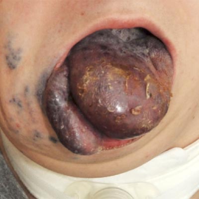
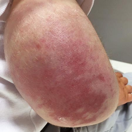
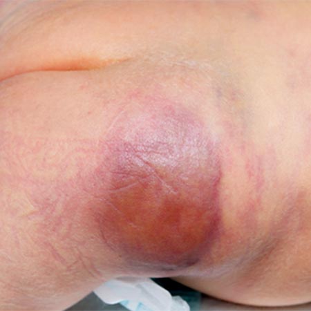
These two types of coagulopathy occur almost exclusively in the clinical pictures of kaposiform hemangioendothelioma / tufted angioma (KHE/TA) as Kasabach-Merritt phenomenon (KMP) and in large-volume venous malformations (VM) as localized intravascular coagulation (LIC), as presented here. The latter can also turn into disseminated intravascular coagulation (DIC) in response to certain triggers such as surgery. Thus, both coagulation phenomena can assume life-threatening severity if inadequately treated or not at all.
Inadequate therapy comes in many forms. When coagulation phenomena progress to bleeding due to pronounced coagulation factor consumption in DIC, the necessary heparin therapy is often applied too late or not at all out of concern for a possible increase in bleeding. If platelet concentrates are used too early or too frequently for deep thrombocytopenia in KMP, massive inflammatory triggering of platelet aggregates can ensue, with massive swelling and inflammatory reaction of the tumor and subsequent worsening of the coagulation situation; in these circumstances transarterial embolization can be life-saving.
Early recognition, differential diagnosis and prophylaxis as well as specific therapy of these coagulation disorders associated with specific vascular anomalies are described in more detail below. In these circumstances, close collaboration with hemostaseologists, laboratory physicians, and anesthesiologists at an interdisciplinary center is very helpful.