Chapter: The classification of vascular anomalies
Article: 7 of 8
Update: June 04, 2021
Author(s): Sadick, Maliha | Wohlgemuth, Walter A.
Vascular tumors are subdivided into benign, locally aggressive or borderline and malignant tumors. They are solid, often progressive vascular tumors, which are based on true cell proliferation. Thus, they do not consist of vascular or sponge-like, fluid-filled cavities like vascular malformations, but form a solid tumor mass. Accordingly, they are neither soft to the touch nor are they compressible. Vascular tumors may be congenital (e.g., congenital hemangioma or kaposiform hemangioendothelioma) or may develop later on tissue that previously appeared healthy.
The main representatives of benign vascular tumors are the very common infantile hemangioma and the small group of congenital hemangiomas; all other relevant vascular tumors are extremely rare. Based on the ISSVA classification, the following table provides an overview of the classification of vascular tumors.
| Biological behavior | Classification |
|---|---|
| Benign vascular tumors |
|
| Locally aggressive and borderline vascular tumors |
|
| Malignant vascular tumors |
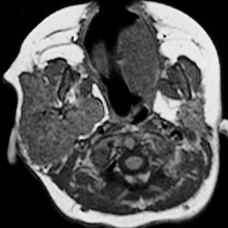
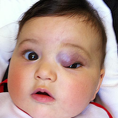
Infantile hemangiomas, based on excessive angiogenesis and endothelial cell proliferation, are the most common benign vascular tumor, accounting for more than 90% of the cases. In the weeks following birth, they become visible as a raspberry-red spot on the skin, which quickly grows larger and larger. Immunohistochemically, they express the Glut-1 marker that confirms the diagnosis. Characteristic features are their rapid proliferation and size increase in the first few weeks to months after birth, followed by a plateau phase with subsequent involution und regression starting from the age of 1 year. In most cases, infantile hemangiomas regress spontaneously without the need for therapy. Infantile hemangiomas can be focal, multifocal and segmental and can be superficial, deep or mixed in cutis or subcutis. A clinical feature of superficial infantile hemangiomas, also known as “blood sponges”, is their raspberry-colored, berry-like appearance with sharp demarcation to the surrounding skin.
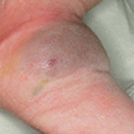
Congenital hemangiomas are very rare benign vascular tumors. In contrast to infantile hemangiomas, they are fully developed at birth and either regress rapidly (rapidly involuting congenital hemangioma, RICH), only partially (partially involuting congenital hemangioma, PICH) or not at all (non-involuting congenital hemangioma, NICH). Postpartum they are no longer growing and are immunohistochemically negative for Glut-1 marker. Unlike infantile hemangiomas, they are not raspberry red but rather bluish livid and may show a pale rim to the surrounding skin.
Other benign vascular tumors can be clinically similar to hemangiomas, so that a clear diagnosis may not be easy during the initial macroscopic inspection. In such cases, patient history (already present at birth?) and indications of rapid expansive or infiltrative growth are relevant criteria for differentiating from locally aggressive or malignant vascular tumors. If the biological behavior of the vascular tumor cannot reliably be assessed clinically and macroscopically and ultrasound also does not enable the findings to be clearly assigned, a biopsy should be performed to safely exclude a malignant (vascular) tumor. Presentation of the patient in a specialized interdisciplinary center for vascular anomalies may certainly be useful in this case.