Chapter: Coagulation disorders
Article: 5 of 6
Update: Feb 24, 2021
Author(s): Wohlgemuth, Walter A.
The very slow-flowing blood in slow-flow malformations (in practice, mainly large venous malformations) also leads to permanent, spontaneous coagulation processes at the site of the pathologically dilated, dysplastic vascular spaces due to the lack of blood flow (stasis). This is called localized intravascular coagulation (LIC) within the venous malformation.
Blood stasis is one of the three classic components of Virchow's triad in coagulation activation. Stagnant blood coagulates. At the same time, activated coagulation also activates local fibrinolysis in parallel and this attempts to break down these clots again to counterbalance overactivation.
In the majority of patients with somewhat larger venous malformations, these processes can also be measured in the blood in the form of a permanently elevated D-dimer level.
The cause of LIC is venous stasis of blood in the abnormal, dilated venous spaces of the venous malformation (VM). This activates plasmatic coagulation. This coagulation activation occurs primarily via activation of factor IIa (thrombin), which amplifies its own formation and activates the formation of linked, insoluble fibrin (F Ia) via activation of factors V, VII, XI from soluble fibrinogen (F I).
Video: Blood stasis within a venous malformation on ultrasound (B-scan). The very slow blood flow in the dilated vascular spaces of the VM results in the formation of small thrombi, which appear slightly more echogenic (light gray) in the sonogram in contrast to normally anechoic blood (completely black). © Wohlgemuth
In addition to stasis of the blood, it is likely that atypical vascular endothelial properties in the dysplastic vessel walls of the venous malformation also play a procoagulatory role.
The presence of permanent LIC with constantly elevated D-dimer levels is described in the literature in 42% to 70% of patients with venous malformations.
Measurably elevated blood D-dimers in VM patients are more likely to occur, depending on the following factors:
The above-mentioned factors correlate positively with continuously ongoing, localized intravascular coagulation.
Almost constantly elevated D-dimer blood levels are the most important laboratory parameter for ongoing LIC in patients with venous malformation (VM). More than one quarter of patients with venous malformations and more than three quarters of patients with large intramuscular venous malformations show constantly highly elevated D-dimer levels (> 1.0 mg/ml). This is true even in normal life without other concomitant reasons for D-dimer elevation such as thrombosis, pulmonary embolism, tumor disease, inflammation, pregnancy, etc.
The mildly elevated D-dimer levels sometimes measurable in other patients without a vascular anomaly, especially with increasing age, ulcerations or wounds, oral contraception or nicotine abuse, are usually much less pronounced than the levels in VM patients. However, these co-factors may be added intercurrently in patients with venous malformations and may further increase their D-dimer levels.
These clearly elevated D-dimer levels in patients with venous malformations, who may be otherwise quite healthy, do not occur in any other disease and are thus an important differential diagnostic component. Stagnant blood within the venous malformation results in classic fluid-fluid levels on imaging due to corpuscular blood components sinking with gravity in the absence of blood flow.
Elevated D-dimer levels in vascular anomaly patients always indicate a venous malformation component: whether as a simple or combined venous malformation (e.g., CVM, CVLM) or in the context of a malformation syndrome such as Klippel-Trénaunay syndrome (CLVM + limb hyperplasia).
However, a non-elevated blood D-dimer value cannot exclude the presence of what will usually be a small venous malformation.
D-dimers are normally not elevated in fast-flow malformations (AVM, AVF), pure lymphatic malformations (without hemorrhage), and even in small glomuvenous malformations.
With increasing LIC, there may also be an initially slight decrease in fibrinogen. Soluble fibrin complexes are then measurable in the blood at mild to markedly elevated levels (+ to +++).
Factor VIII-vWF complexes could be detected in up to 80% of patients with major venous malformations.
Decreases in factor VIII and vWF < 60% are found in approximately one third of patients with venous malformations.
The number of platelets in the blood is usually normal or at most slightly decreased in LIC, unlike in patients with Kasabach-Merritt phenomenon (KMP).
These coagulation events are a lifelong, ongoing process in VM patients, i.e., independent of acute episodes of disease or recent thrombophlebitis. They are detectable throughout life even in completely healthy patients with large venous malformations.
At the same time as the ongoing localized coagulation (LIC), there is also reactive secondary hyperfibrinolysis, which counterbalances the ongoing coagulation activation. This is also easily measurable in the laboratory by means of ROTEM analysis or measurement of TGT (thrombin generation time).
These two parallel processes (simultaneous coagulation activation and fibrinolysis) lead to constantly increased turnover, consumption and replenishment of coagulation factors. If the replenishment of these factors is slower than the consumption, this can be measured by a decrease in fibrinogen, factor XIII and antiplasmin in particular. This may be an indicator of imminent disseminated intravascular coagulation (DIC).
In patients with large venous malformations (VM), the continually ongoing LIC can turn into disseminated intravascular coagulation (DIC) as a result of further overactivation. DIC equates to potentially life-threatening coagulation failure.
A permanently elevated D-dimer level (> 1.8 mg/ml) and an already low-normal or even low fibrinogen level (< 100 mg/dl) as well as the clinical occurrence of spontaneous bleeding are warning signs for the occurrence of DIC. Low fibrinogen indicates imminent danger.
If LIC turns into DIC, e.g., during open surgical resection of a VM, massive intraoperative bleeding may occur. This is caused by thrombocytopenia, consumption of coagulation factors and fibrinogen, which can hardly be replenished or substituted quickly enough. This is intensified by the simultaneous anticoagulant effect of fibrin degradation products and the consumption of hyperactivated platelets.
In parallel, disseminated intravascular microthrombosis may occur in the setting of DIC.
Typical risk situations that can lead to the triggering of DIC in patients with VM are all procoagulatory situations:
Patients with large venous malformations and the following laboratory constellation are particularly at risk:
In these situations prophylactic anticoagulation is necessary before any intervention/surgery.
The extent of continually ongoing LIC and thus clot formation in parts of the venous malformation also correlates well with the formation of local, often painful thrombophlebitis within the venous malformation. This pain is triggered by constant ongoing formation of microthrombi and concomitant activated fibrinolysis.
Some of these continually forming blood clots are larger, which means they are only slowly cleared via local inflammatory processes of fibrinolysis. These larger clots lead to the formation of painful thrombophlebitis within the venous malformation.
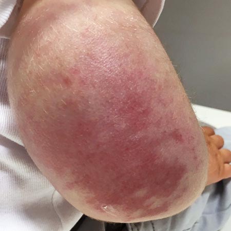
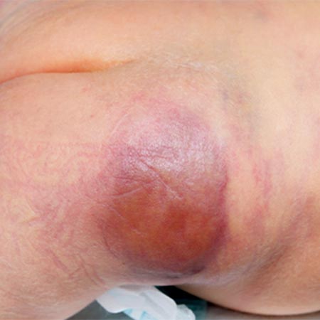
Clinically, there is local pain, local induration and swelling. If thrombophlebitis is superficial on the skin, local redness is often visible as well. The duration of this circumscribed inflammation within the venous malformation ranges from a few hours to 3 to 5 days, sometimes longer. D-dimer levels are further elevated during such thrombophlebitis.
The normal organizing degradation of larger local thrombi or thrombophlebitis can lead to collagen deposition, which is palpable as a circumscribed induration over a longer period of time. If the thrombus then still cannot be completely degraded in the venous malformation, calcium deposition and increasing local, shell-like or popcorn-like calcification will occur over time. The final form is the calcified phlebolith as a round calcified lesion 1 cm in size on X-ray images of the venous malformation.
The painful inflammatory processes associated with spontaneous LIC and secondary hyperfibrinolysis or thrombus formation and degradation within the venous malformation explain typical clinical pain phenomena in VM patients.
The basic mechanism is that the pain worsens pain with increased clot formation and improves with less clot formation or improved clot degradation.
Thus, there is a typical worsening of pain during immobility or after prolonged lying in bed. The cause is increased clot formation due to increased blood stasis. Another typical worsening of the pain is found in the second half of the night or in the early morning before a person gets up or makes their first movement (with pumping and evacuation of the venous spaces). Lying motionless at night, similar to immobility, leads to increased blood stasis and thus LIC in the venous malformation. Frequently, this pain is consequently improved by active movement of the affected body regions (so-called “start-up phenomenon”). This involves pumping out the affected vascular spaces via the muscle pump and flushing them free of small clots.
In venous malformation patients, the described mechanism also leads to a worsening of pain within the venous malformation due to general clot activation in the context of inflammation, infection, wounds or surgery. The same is true in cases of fever, thirst/exsiccosis, and long all-nighters associated with excessive alcohol consumption (exsiccosis due to the diuretic effect of ethanol). Venous malformation patients should avoid such situations if possible.
The localization of the venous malformation in the body is also important for the degree of painfulness. If unfavorably located near peripheral nerves or more pain-sensitive tissues (such as at the periosteum of the bone or fingers and toes), even small venous malformations may be significantly painful. The continually ongoing coagulation processes lead to local inflammation near these pain-sensitive structures.
The most important conservative therapy is appropriate compression therapy, as this directly decreases LIC via volume reduction of the affected dysplastic vascular spaces of the venous malformation. Smaller VM volume means less LIC in the malformation. However, not all body regions are amenable to compression measures.
Sufficient periods of exercise decrease the stasis of blood in the venous malformation. Therefore patients with large venous malformations, especially of the extremities, should never sit or stand for more than a maximum of one hour without intermittent exercise.
Elevation of an affected limb not only relieves pressure (and decreases volume), but also causes blood to drain away (thus decreasing microthrombi) with subsequent refilling by unclotted, fresh blood. This serves to prevent the formation of large thrombi and decrease LIC.
Local cooling is usually soothing and comfortable for acute painful inflammation and pain associated with acute thrombophlebitis. Via local vasoconstriction during cooling, the vascular volume also decreases and so does the clotting burden. Unfortunately, the capacity for vasoconstriction in the dysplastic vessel walls of a venous malformation is often limited due to dysplasia or lack of smooth muscle cells in the vessel wall of the venous malformation.
In addition, coagulation-inducing situations such as oral contraception, tobacco smoking, dehydration, immobility, etc., should be avoided.
The basis of drug therapy for LIC in venous malformations is mild inhibition of plasmatic coagulation. Classic therapy is the administration of a low-molecular-weight heparin (LMWH) at a prophylactic dosage. Low-molecular-weight heparins are also very effective for prolonged, severe pain and thrombophlebitis, e.g., once daily for 5 to 7 days in prophylactic doses.
Special consideration should be given to prophylactic heparin administration before major interventional or open surgical procedures for venous malformations. In the case of abnormal coagulation laboratory findings, extensive LIC and large-volume venous malformation, heparin should be administered at least 3 to 5 days before a procedure to prevent the possible progression of LIC into DIC. The same dose of LMWH should be continued for at least 20 days after a procedure.
The new or direct oral anticoagulants (DOACs), as anti-Xa inhibitors based on an analogous mechanism of action to heparin, are also clinically very effective in thrombophlebitis treatment and prophylaxis as well as LIC reduction. However, larger studies on this are not yet available, and its use remains off-label. The advantages are that a relatively low dose is required and the oral route of administration can be used. Case study experience with rivaroxaban and apixaban in prophylactic doses showed good clinical efficacy.
Vitamin K antagonists (e.g., warfarin, phenprocoumon) are also effective in pain control, but inferior in overall profile regarding risk-benefit ratio because of the risk of bleeding. In cases of thromboembolism through communicating veins of a venous malformation with the deep conducting vein system, especially in conditions following pulmonary embolism, these communicating veins should be closed by intervention or surgery. Permanent anticoagulation may be indicated if this is not possible.
Antiplatelet agents such as ASA or clopidogrel are less effective in venous malformations, both clinically and for pathophysiologic reasons. They inhibit platelet aggregation instead of plasmatic coagulation, which plays a much smaller role in LIC within the venous malformation, in contrast to the Kasabach-Merritt phenomenon.
Cortisone, interferon, or propranolol are not effective or useful in this context.
All invasive methods that reduce the volume of the venous malformation naturally lead to an improvement of LIC within the venous malformation as well as thrombophlebitis-associated pain via a reduction of the stagnant blood volume.
Thus, reducing the volume of the venous malformation by sclerotherapy is the first choice in further invasive therapy. Other local ablation procedures (such as laser therapy, radiofrequency ablation, microwave ablation, cryoablation) can also be applied in this regard with the aim of reducing the volume of the dilated vascular spaces of the venous malformation.
The use of open surgical resection procedures alone is reserved for specialized centers because of the mostly very extensive infiltrative lesions and the risk of activation of coagulation with conversion of a LIC into a DIC. Catastrophic coagulation failure is possible.
Of course, combinations of the above-mentioned invasive therapeutic procedures are also possible, for example, first sequential sclerotherapy followed by resection of the reduced VM compartments.
The importance of starting heparin administration as DIC prophylaxis 3 to 5 days before major invasive procedures in the case of conspicuous coagulation laboratory findings with extensive LIC and large venous malformation must be stressed once again.