Author(s): Kertai, Michael Amir
Author(s): Kertai, Michael Amir
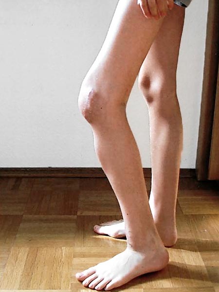
Left knee in maximum active extension possible. The pre-patellar scar indicates a previous attempt to remove the tumor.

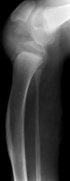
Lateral X-ray of the left knee joint with osseous dysplasia of the upper and lower leg. The tibia has assumed a curvature in this area during growth. The fibula, on the other hand, is straight. Nevertheless, the essential problem is limited to the knee joint.

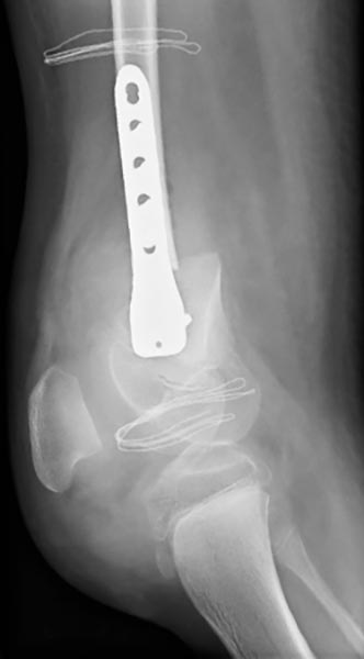
As a result of surgical alignment of the axis of the femur, the knee joint could be extended immediately postoperatively. Fixation by means of osteosynthesis. The leg length discrepancy was compensated by simultaneous removal of a bone wedge.

This 12-year-old patient with PTEN hamartoma syndrome developed a progressively growing, painful PTEN hamartoma in his thigh area directly above the kneecap. As a result of the pain caused by the mechanical pressure during knee extension (patella pressing against the tumor), among other things, he constantly adopted a permanent knee flexion posture to avoid pain. Active knee extension was also no longer possible during the course of the disease owing to the abnormal osseous growth of the femur.
In addition, the patient had leg length discrepancy with an excess length of about 3 cm for the left, affected leg.
Published: 2018
All images © Kertai


