Chapter: Wounds and ulcerations
Article: 4 of 7
Update: Mar 10, 2021
Author(s): Ott, Hagen
The skin defect and other possible influencing factors, such as wound secretion, crust formation or necrosis, promote the penetration of pathogens into the wound (contamination). If proliferating bacteria can be detected in the wound, this is referred to as “colonization”, which is regularly present in chronic wounds.
The term “critical colonization” is used when the bacterial count is so high that it delays healing of the wound or makes it impossible (“bacterial overload”). A limit of more than 105 microorganisms per mm3 is frequently specified for this critical colonization.
Not until the patient develops an immune response is the situation called an “infection”. This may be a local infection confined to the wound area and adjacent structures or it may lead to a systemic immune response.
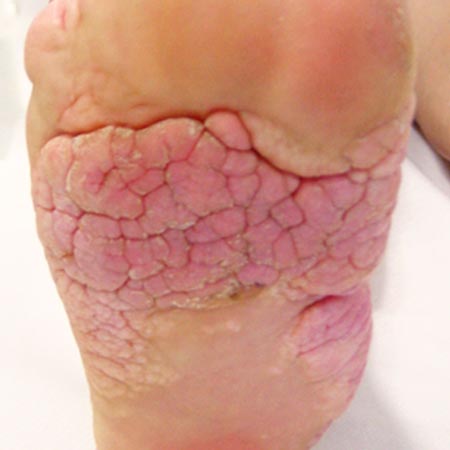
Local symptoms of wound infection are (selection):
A systemic infection must be assumed if fever, chills, generalized lymph node enlargement and/or deterioration of the patient’s general condition occur.
A bacteriological skin swab should be ordered if a secondary infection is suspected, if the wound fails to heal or if the symptoms increase again after wound healing has already begun. In these cases, the swab should be taken from the depth of the wound and not from superficial, crusty areas in order to avoid erroneous cross-contamination with physiological skin germs. The sample is preferably taken in a spiral from the outside to the inside in order to obtain a representative sample.
If critical colonization or localized infection is suspected, the wound is first cleansed with sterile, physiological saline or Ringer’s solution. This is followed by local antiseptic treatment primarily with octenidine or polihexanide, which are available as ready-to-use medication and as individual formulations in different galenics. Development of resistance to these substances has not so far been described and they have practically no allergenic potential.
Polihexanide is even thought to have a wound-healing effect, so that in many places it is considered the topical antiseptic of first choice. Octenidine is commonly used to treat acute, bacterially contaminated wounds at a concentration of 0.1%, while polihexanide at concentrations of 0.02% and 0.04% is preferred for the treatment of chronic wounds, both prophylactically and in cases of colonization and infection, respectively. When using octenidine dihydrochloride, it should be noted that it must not be injected into puncture channels or other “closed” wounds as a result of increased pressure because of the risk of prolonged tissue damage.
Topical antibiotics such as neomycin should not be used for local therapy of wounds due to the increasing development of resistance and the risk of contact allergy.
If there is clinical and/or laboratory evidence of a systemic reaction, oral or intravenous antibiotic therapy should be initiated early, based on the resistogram.