Chapter: Wounds and ulcerations
Article: 3 of 7
Update: Mar 10, 2021
Author(s): Ott, Hagen
In patients with vascular anomalies, wounds require an additional, detailed patient history, which should include the following questions:
As a second step, affected patients are examined thoroughly and the findings are ideally entered on a wound documentation form. In addition, standardized photographic documentation is indispensable, especially in the case of chronic wounds.
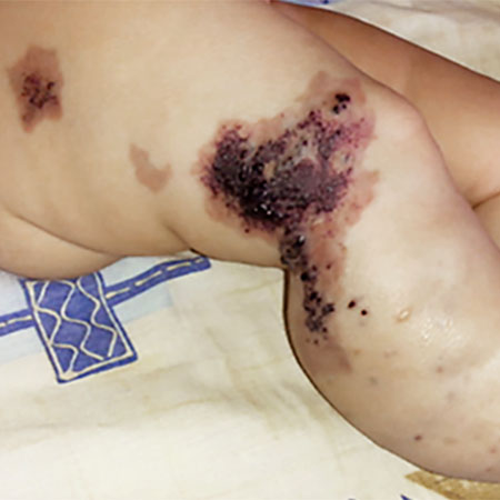
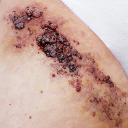
First, the location and dimensions of the wound are recorded. Particular attention should be paid to the wound margin, which may be undermined, edematous or macerated. The wound bed should also be inspected. Depending on the wound phase, it may be yellowish with fibrin (exudative phase), show red granulation tissue (proliferative phase) or already show renewed closure by epidermal cells (reparative phase).
In addition, the extent and nature of wound secretions are documented and it is checked whether clinical signs of a secondary infection are present. Dry, black or avital, whitish wound areas indicate dead tissue and are referred to as necrosis. They are often surrounded by an inflammatory marginal wall and are deficient in blood supply, thereby preventing problem-free wound healing.