Author(s): Wohlgemuth, Walter A.
Author(s): Wohlgemuth, Walter A.
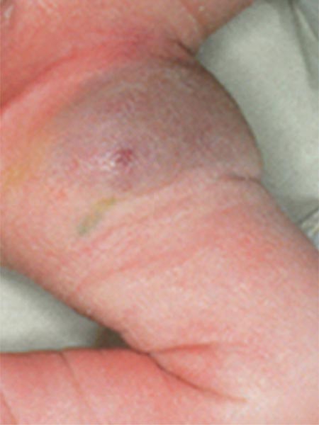
Boy with congenital, blue-livid discolored tumor in the left groin and proximal thigh (photograph from day 3 of life).
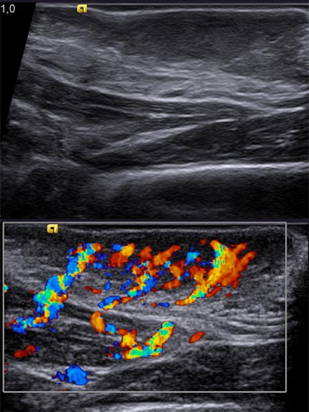
On B-scan ultrasonography (top image), the tumor is relatively homogeneous, hypoechoic, and clearly solid, not compressible. Color-coded duplex ultrasonography (bottom image) shows intense perfusion via multiple arterial vessels. This is suggestive of a congenital vascular tumor and, in this case when combined with the bluish appearance, the special case of a congenital hemangioma.
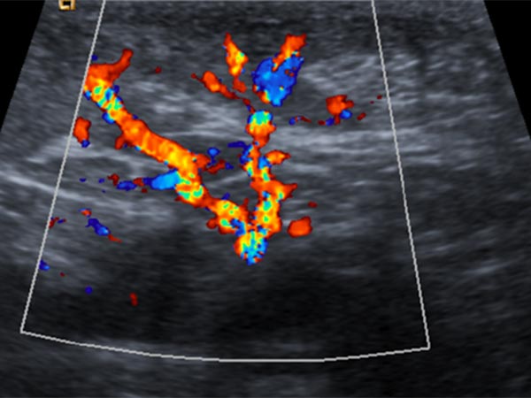
Control sonography (color-coded) at 4 months of age shows no change in echogenicity, especially no signs of involution. Continued strong perfusion and no increase in echogenicity, as would be the case with a rapidly involuting congenital hemangioma (RICH).
Photograph taken at the end of the 4th month of life. Both the expansion and the volume show no signs of involution.
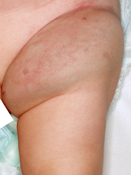
Photograph at the end of the 6th month of life, still no significant changes, the vascular tumor appears rather increased in size.
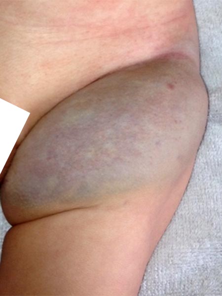
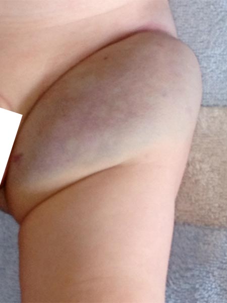
End of the 10th month of life, still no signs of involution. RICH (Rapidly Involuting Congenital Hemangioma) can thus be safely excluded. Sonography also shows no change. Blood coagulation and platelets without pathological changes, no Kasabach-Merritt phenomenon is present. The diagnosis of NICH (Non-Involuting Congenital Hemangioma) is thus the most probable.
Coronal T2-weighted, fat-saturated MRI shows the tumor in the left groin as homogeneous and highly hyperintense (13 months of age) and clearly solid. Incidental findings include the soaked diaper, also with high signal intensity.
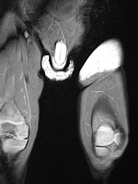
Same MRI sequence, coronal slice. The tumor is not only epifascial, but also shows a small subfascial extension under the fascia lata into the gluteal muscles. Thus, clearly infiltrative behavior.
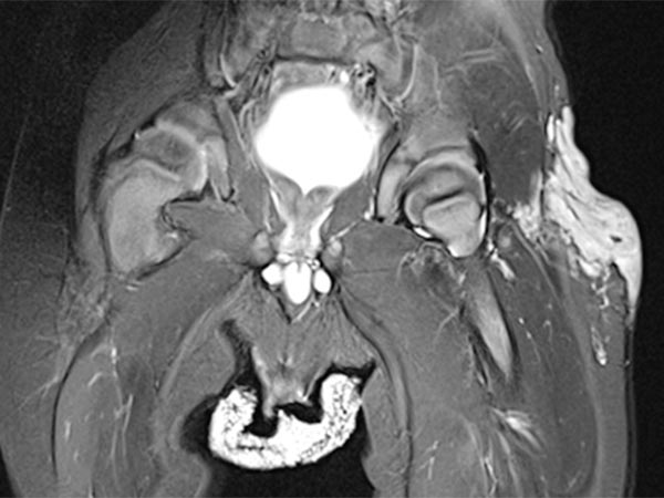
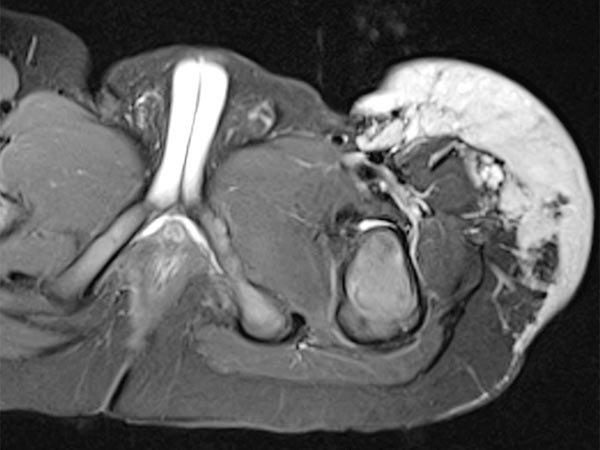
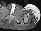
Axial slice of MRI (T2-weighted, fat-saturated) also clearly shows the infiltration of the musculature by the mass. Hemangiomas as vascular tumors can also exhibit such infiltration without necessarily being malignant.
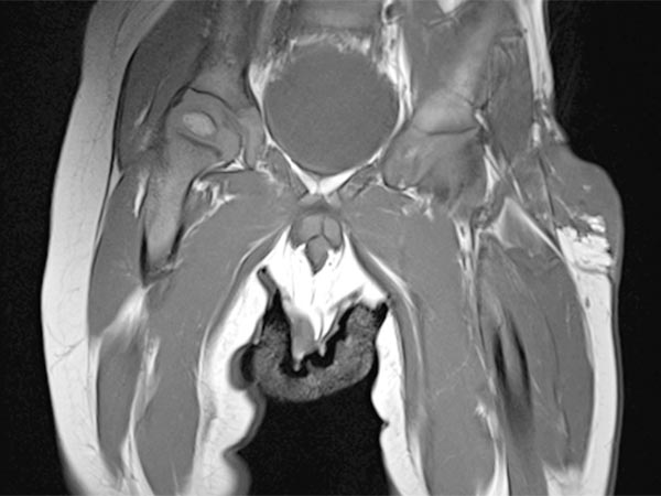
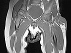
In non-enhanced coronal T1-weighted MRI, the tumor is isointense to the musculature without containing adipose tissue. Thus, it is hardly distinguishable from the musculature in this sequence.
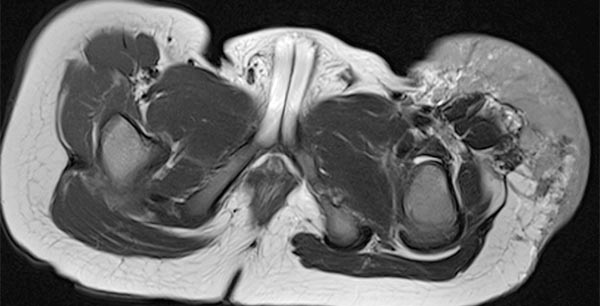
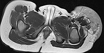
Axial T2-weighted MRI without fat saturation shows the tumor as homogeneous and only slightly hyperintense. It is more hyperintense than muscle, but overall much less hyperintense than the surrounding subcutaneous adipose tissue.
Coronal T1-weighted, fat-saturated MRI after contrast administration. The tumor shows homogeneous, strong enhancement. Inside it, two flow voids as a sign of strong arterial perfusion.
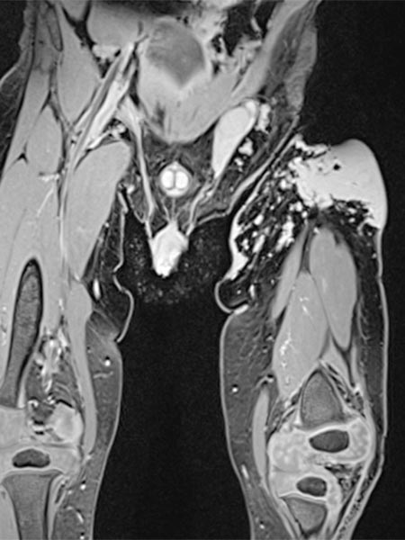
Contrast-enhanced dynamic MR angiography (coronal plane).
30 s after contrast injection in the early arterial phase, there is immediate early enhancement of the tumor in the left groin.
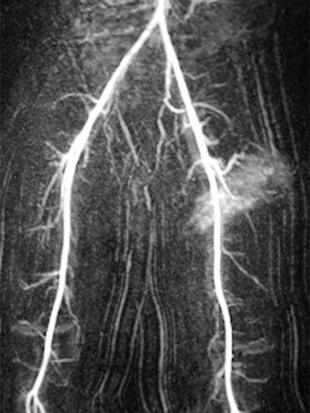
Contrast-enhanced dynamic MR angiography (coronal slice orientation).
41 s after contrast injection in the late arterial phase, there is further, rather diffuse, strong enhancement of the tumor ("tumor-blush") in the left groin, corresponding to a solid vascular tumor.
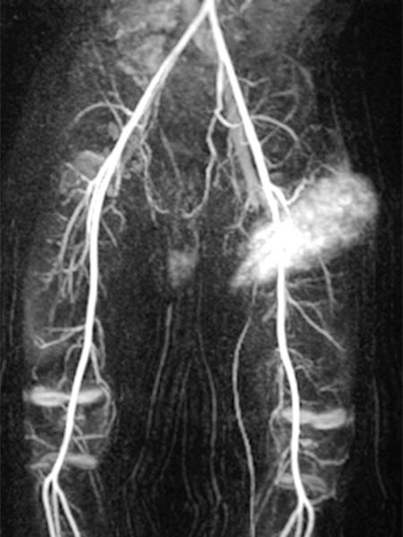
Contrast-enhanced dynamic MR angiography (coronal slice orientation).
57 s after contrast injection in the early venous phase, the entire tumor continues to enhance strongly. The image now also displays a dilated early drainage vein in comparison of sides. The venous drainage (left iliac vein) has dilated because of the strong tumor perfusion with increased venous outflow.
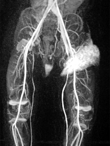
Contrast-enhanced dynamic MR angiography (coronal slice orientation).
130 s after contrast injection in the late phase, the strong enhancement of the tumor in the left groin remains, no early "wash-out". Additionally, the enhanced venous drainage via the left iliac veins still contrasts.
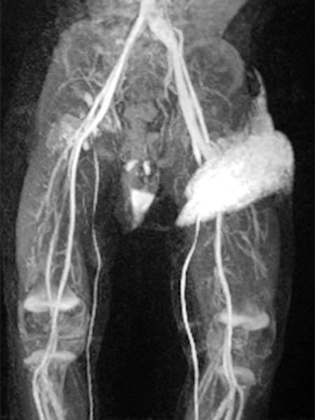
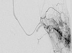
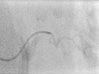
As there was still no tendency to regression of the tumor at the end of the 3rd year, embolization was performed after biopsy to induce involution. Digital subtraction angiography (DSA) shows a microcatheter superselectively introduced into a tumor vessel. The tumor is heavily perfused and lobulated, very early venous outflow, typical of an NICH.
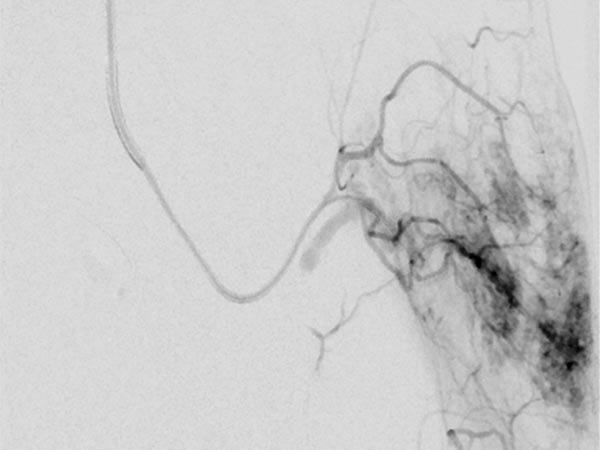
Particle embolization with spherical particles 250 micrometers in size via the microcatheter inserted superselectively into the tumor.
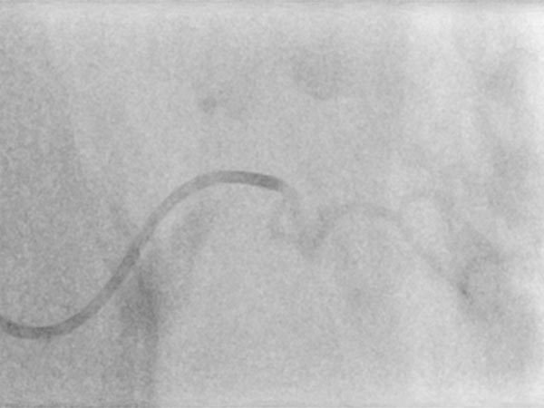
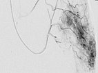
More tumor vessels with blush-like diffuse enhancement, typical of a vascular tumor / NICH. All these vessels must be selectively embolized to induce involution.
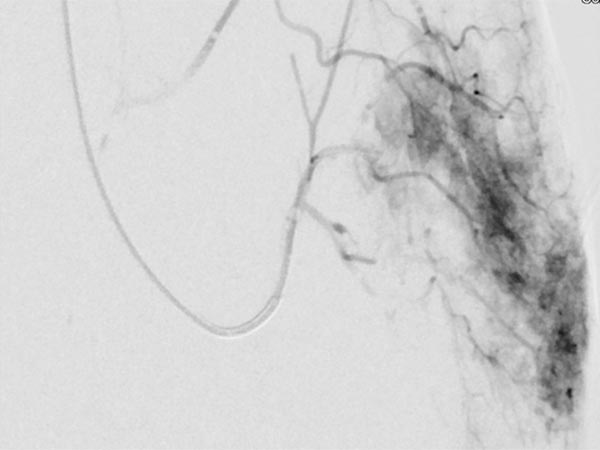
Further selective particle embolization. The embolization particles mixed with contrast medium remain in the tumor vessels.
![Non-involuting congenital hemangioma [Translate to English:] Embolization: contrast medium remain in the tumor vessels](/fileadmin/images/patientenbeispiele/29-nich-leiste/f29-20-non-involuting-congenital-hemangioma-embolisation.jpg)
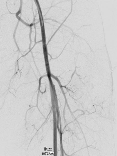
Overview angiography via the left external iliac artery also shows no remaining perfusion of the tumor anymore, thus the tumor vascularization is successfully and superselectively completely occluded. All non-pathological arteries are preserved.
Immediately after embolization, the color of the tumor became much paler, and the volume also decreased significantly as a sign of involution induced by embolization with elimination of vascularization.
An initially planned, additional surgical resection of the tumor remnants was abandoned because of the good result. The further course is awaited.
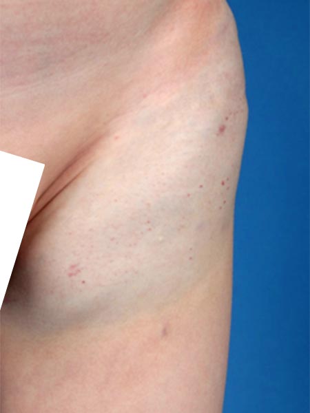
In the further course after embolization, the tumor has greatly decreased in volume, it is now no longer raised and is asymptomatic.
Photograph at the age of 4 years 8 months. The only residual NICH is a local bluish discoloration of the skin.
Further invasive therapy is no longer necessary.
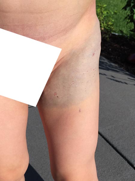
Photograph of the patient at the age of 7 years and 10 months.
The NICH is still visible through the skin with a bluish shimmer, but hardly shows any more space-occupying effect and is less color-intensive.
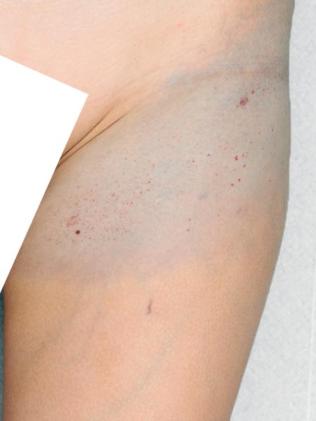
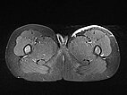
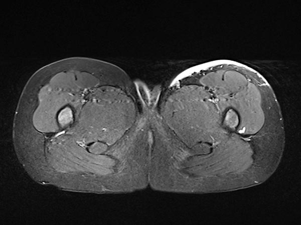
Axial, T2-weighted, fat-suppressed MRI of the patient at 7 years and 10 months of age.
The NICH is still visible under the skin as a hyperintense (white) flat structure, but significantly smaller than before embolization.
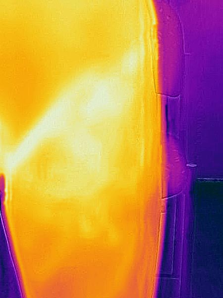
Infrared thermography image of the patient at the age of 8 years and 10 months.
Warmer areas are color-coded lighter to whitish in this image.
The warmer temperature in the skin folds of the groin is normal.
Skin in the area of the residuum of the NICH is still slightly warmer, but not pronounced.
Congenital hemangiomas are very rare vascular tumors and, in contrast to infantile hemangiomas, are congenital, i.e., already fully developed at birth. They usually either shrink rapidly and spontaneously within a few months (RICH) or do not involute at all (NICH). They do not respond to the administration of propranolol. For extensive tumors in unfavorable locations, superselective embolization after biopsy to induce involution may be an alternative to open resection in individual cases. The resection planned for this patient could be omitted due to the good embolization result.
Published: 2020
All images © Wohlgemuth






![Non-involuting congenital hemangioma [Translate to English:] Embolization: contrast medium remain in the tumor vessels](/fileadmin/_processed_/3/2/csm_f29-20-non-involuting-congenital-hemangioma-embolisation_1f7261168b.jpg)


