Chapter: Imaging
Article: 3 of 10
Update: Feb 05, 2021
Author(s): Müller-Wille, René
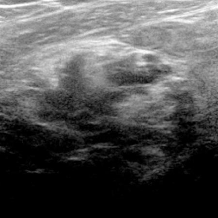
Sonography is a non-invasive and radiation-free, harmless imaging procedure using ultrasound waves.
The sound waves are generated in the ultrasound transducer by means of the piezoelectric effect and the returning waves reflected differently by different tissues are detected. For the imaging of superficial lesions, linear ultrasound probes with high frequencies are most suitable. The abdomen and deeper structures are usually examined with convex transducers of lower frequency and thus higher penetration depth. The reflections of the ultrasound waves can be converted in real time into a grayscale, two-dimensional image, the so-called B-mode image.
Structures with low echogenicity are imaged dark (e.g., water) and structures with high echogenicity (e.g., thyroid) are imaged bright (white). In addition to the anatomical structures recognizable on B-mode imaging, the blood flow velocity within the vascular system can be precisely measured on the basis of the Doppler effect (pulsed wave / PW Doppler) and displayed in two dimensions (color Doppler, color-coded duplex sonography). A further development is contrast-enhanced ultrasound (CEUS), in which intravenously applied microbubbles enable the perfusion of vessels and tissues to be assessed.
Ultrasound elastography allows for imaging of the hardness / softness of a tissue in color.
| Mode | Description | Application |
| B-mode (brightness mode), 2D real time | Two-dimensional, grayscale images | Characterization and extent of the lesion Guidance of percutaneous therapy |
| PW Doppler | One-dimensional measurement of blood flow velocity | Differentiation between slow-flow and fast-flow vascular malformations |
| Color-coded duplex sonography (CCDS) | Two-dimensional, color-coded imaging of the blood flow velocity | Differentiation between slow-flow and fast-flow vascular malformations |
| Contrast-enhanced sonography (CEUS) | Imaging of contrast medium using microbubbles | Differentiation between slow-flow and fast-flow vascular malformations as well as tissue perfusion |
| Elastography | Color-coded imaging of tissue hardness/softness | Differentiation of therapy-induced effects, differential diagnosis |
Sonography is an excellent method for the initial examination when a vascular anomaly is clinically suspected. Especially superficial lesions can be detected very well with two-dimensional B-mode sonography. The clinically important differentiation between slow-flow and fast-flow vascular malformations is thus possible in many cases using color-coded duplex sonography (CCDS) and contrast-enhanced sonography (CEUS). B-mode sonography also serves as image guidance during percutaneous invasive therapies.
A disadvantage of sonography is the relatively small field of view and the often low penetration depth of the ultrasound waves. Therefore, deep lesions cannot be adequately visualized with this modality. Structures that lie behind bone or air cannot be imaged at all. Furthermore, the experience of the examiner also plays an important role in image interpretation.