Author(s): Hülsemann, Wiebke
Author(s): Hülsemann, Wiebke
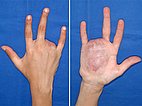
At birth a lesion was found on the ring finger. An “angiodysplasia with extensive shunts” was diagnosed and, at the age of 3 years, the ring finger was amputated because of recurrent bleeding from an ulcer at the fingertip. These photographs were taken at age 12. At that time, intermittent pain was present in the right palm. Reddish, hyperthermic mass in the palm with venous ectasia on the hand, palpable arterial pulse and thrill. Accompanied by soft, elastic palmar swelling extending to metacarpus and hypothenar.
The patient is right-handed.
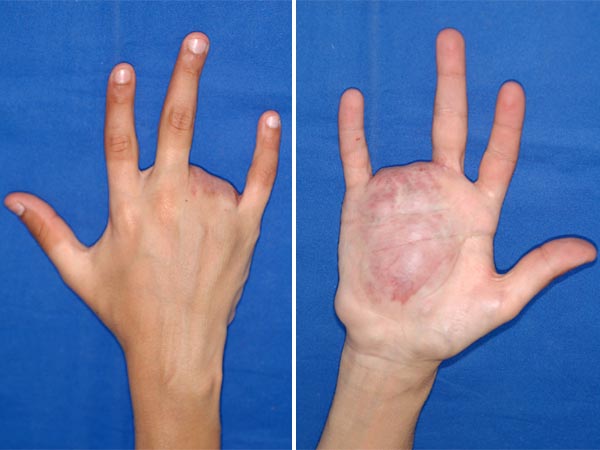
At that time, sonography revealed increased blood flow velocity, multiple vascular channels in the palm, extending into the intermetacarpal space, the ulnar wrist area and including the distal forearm. No signs of left ventricular dilatation on chest X-ray and echocardiography. Pain medication as therapy. A custom-made compression glove without compression of the fingers brought relief. Angiography and transarterial embolizations were performed six times over a one-year period. A partial resection of the embolized compartments of the lesion was performed afterwards.
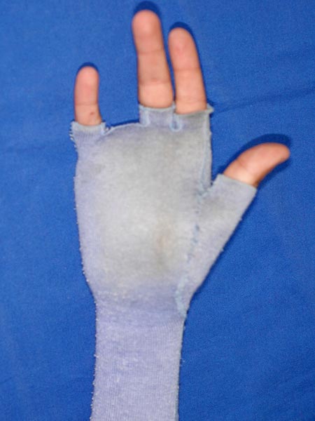
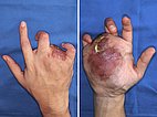
At the age of 18 the situation worsened with a massive increase in pain and multiple relevant arterial hemorrhages in the former amputation area due to an ulcer. Fixed flexion contractures of the middle and little finger. At this time the functionality of the hand was massively limited. A partial amputation of the ulnar part of the hand including the third to fifth ray was now planned. Intended coverage of the resulting defect with soft tissues from the dorsum of the hand.
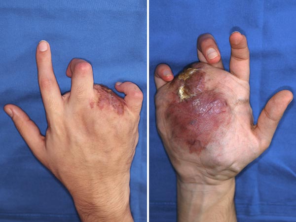
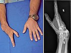
Afterwards clinical stabilization with significant improvement in quality of life. This photo was taken at the age of 25. Initial phantom pain decreased spontaneously from once a week to once every 2 months. The patient was again active in sports with kickboxing and swimming. Completed job training as a mechanical engineer. He now permanently wears a compression glove. On the X-ray (right), parts of the old embolic agent are still visible as radiopaque (white) cast which had occluded the former AVM.
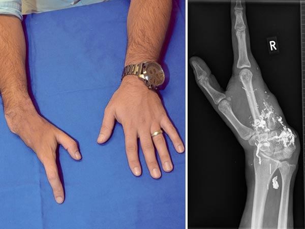
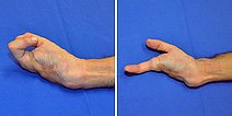
In the 25th year of life, the patient continues to write with his right hand. Weakened grasping with the thumb and index finger of 1 kg on the right side compared to 7 kg on the left.
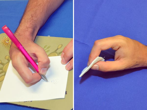
However, unrestricted movement of the remaining digits was found except for a slight limitation in index finger extension due to flexor tendon shortening. Normal sensitivity. No evidence of recurrence at this time.
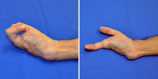
In this patient with an AV malformation of the right hand, arterial bleeding from ulcers of the ring finger led to ring finger amputation at the age of 3 years. After a symptom-free interval with proportional growth, the signs and symptoms increased again markedly during adolescence. A compression glove did reduce pressure and provided some relief. Multiple embolizations at age 16 years and a partial amputation did not have a lasting effect. Emergency treatment was required several times because of recurrent arterial bleeding from the stump area with relevant blood loss. A partial amputation of the entire ulnar hand finally stabilized the situation.
The patient today describes no complaints except for occasional phantom pain. He plays sports without fear and has successfully completed an engineering degree. He continues to write with the affected right hand and continues to wear a compression glove.
Such a rapidly progressive course early in childhood and during puberty is rare but has a high chance of recurrence.
Published: 2018
All images © Hülsemann
