Author(s): Meyer, Lutz
Author(s): Meyer, Lutz
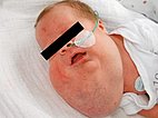
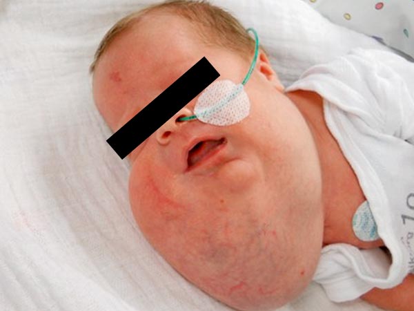
Photograph immediately after birth of a girl with massive cystic lymphatic malformation (LM) of the face and neck. The following description is a typical course of treatment for a patient with such an extensive finding.
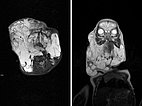
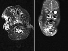
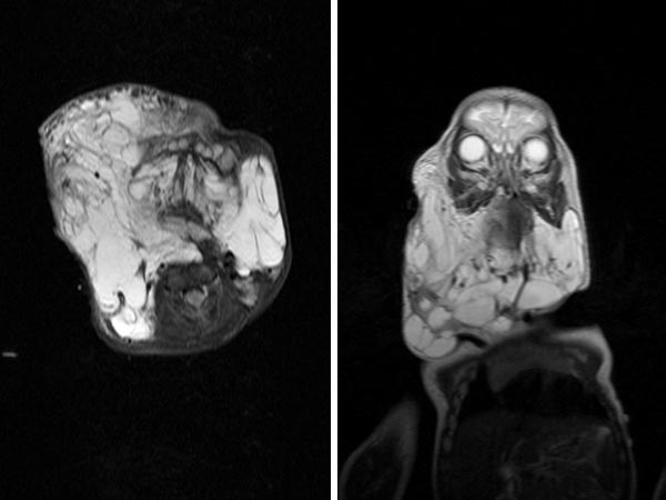
T2-weighted, fat-saturated MRI at 2 months of age shows the full extent of microcystic and macrocystic lymphatic malformation on axial image (left) and coronal image (right).
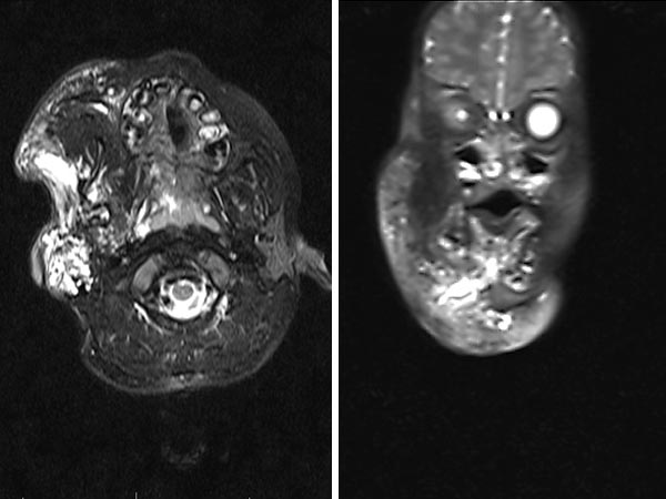
The course of treatment so far: from 2012 to 2013: two open surgical resections in the soft tissue in the submandibular region and first sclerotherapy treatment with OK-432. In 2014: renewed open resection of lymphatic malformation parts of the right cheek via facelift access with neuromonitoring of the facial nerve, combined sublingual resection, 2x sclerotherapy with OK-432. In 2015: CO2 laser resection of lymphatic malformation parts in the pharynx from intraorally. In 2016: closure of the tracheostoma, renewed sclerosing treatment with OK-432. In 2017: 3x sclerosing treatment with bleomycin (10 mg, 15 mg, 15 mg), the last time combined with OK-432. In 2018: last open resection in submental and submandibular region on the right side, thus soft tissue treatment now completed for the time being. The image shows the last MRI in January 2017.
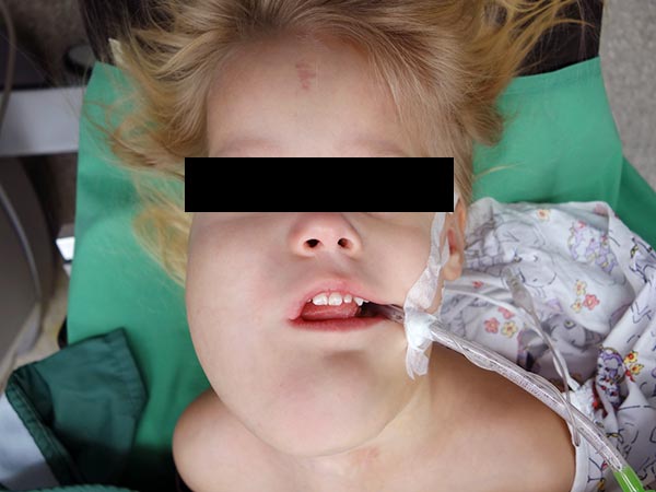
January 2017: Intraoperative image before resection.
Photographs 14 months later, after completion of soft tissue treatment.
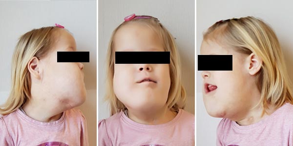
Extensive lymphatic malformations are often accompanied by osseous overgrowth of the jaw and facial bones, with overgrown bone portions that partially protrude at an angle to the opposite side. This causes twisting around the longitudinal axis and is called facial scoliosis.
In relation to this, dental malocclusions can occur, such as an open bite in which the incisors no longer make contact with each other.
Before completion of growth, reduction surgery is possible for an overgrown chin.
After completion of growth, maxillofacial osteotomies can be used to correct the malocclusion.
Published: 2018
All images © Meyer
