Author(s): Meyer, Lutz
Author(s): Meyer, Lutz
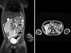
The first case is a 5-year-old girl with distended abdomen. On coronal and transverse T2-weighted, fat-suppressed MRI, an extensive macrocystic lymphatic malformation is visible with large cysts displacing the bowel.
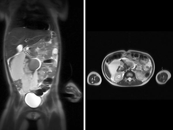
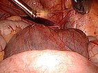
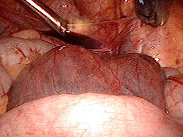
Image during laparoscopic resection: view from the umbilicus to the lower abdomen with some free fluid on the bladder. A large retroperitoneal cyst (lymphatic malformation) bulges in front.
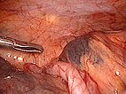
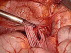
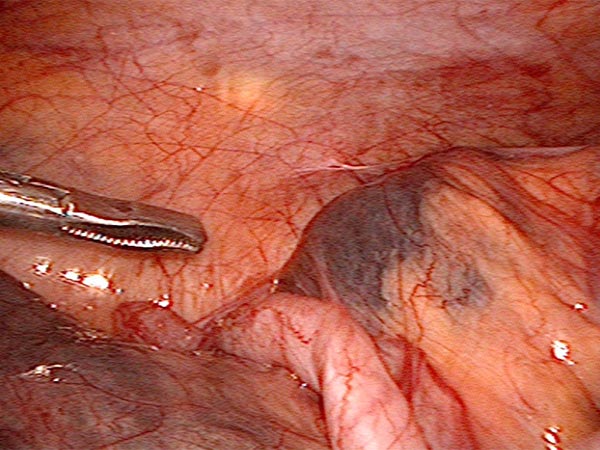
View to the right lower abdomen. Grasping forceps pointing to a cyst in front of the cecum and appendix.
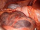
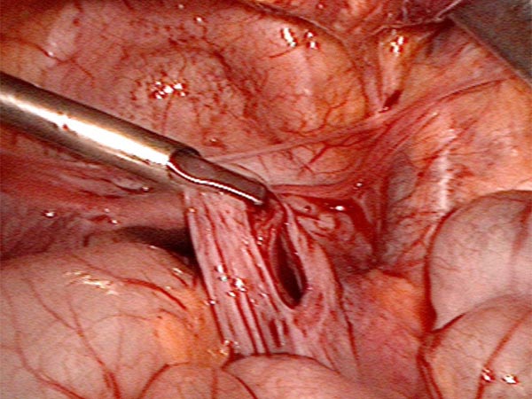
View from the umbilicus to the right upper abdomen with liver and gallbladder. In the mesentery of the small intestine, additional translucent cysts of the lymphatic malformation (LM).
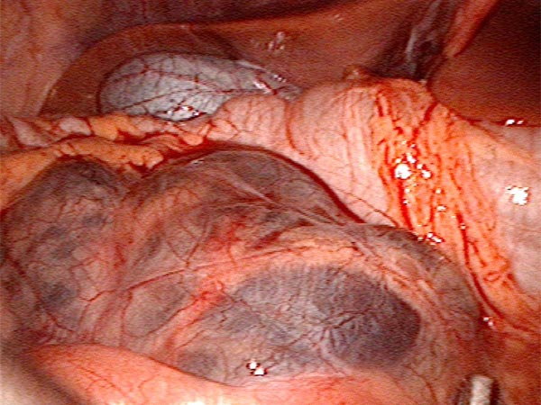
The peritoneum to the posterior abdominal wall is opened and all the cysts are gradually dislodged and removed.
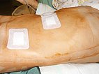
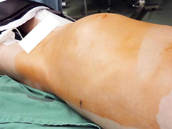
Flat stomach a few days after surgery. No recurrence to date.
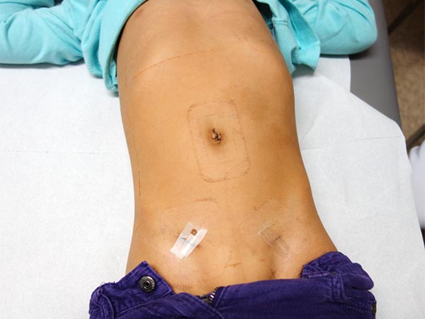
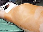
The second case also describes an intraabdominal lymphatic malformation (LM), this time in an 8-year-old boy. The preoperative photograph shows a bulging abdomen.
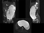
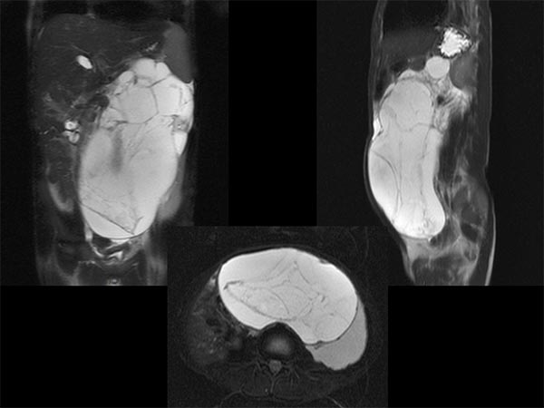
Fat-saturated, T2-weighted MRI in three planes reveals the cause: retroperitoneally located giant cysts due to a macrocystic lymphatic malformation.
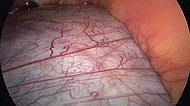
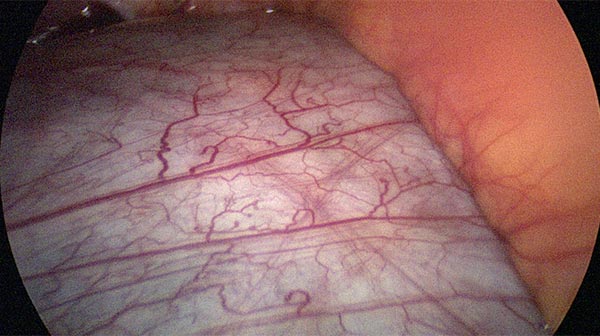
Laparoscopic resection of more than 90% of the cysts. View of the retroperitoneally located cysts of the lymphatic malformation which are visible through the peritoneum.
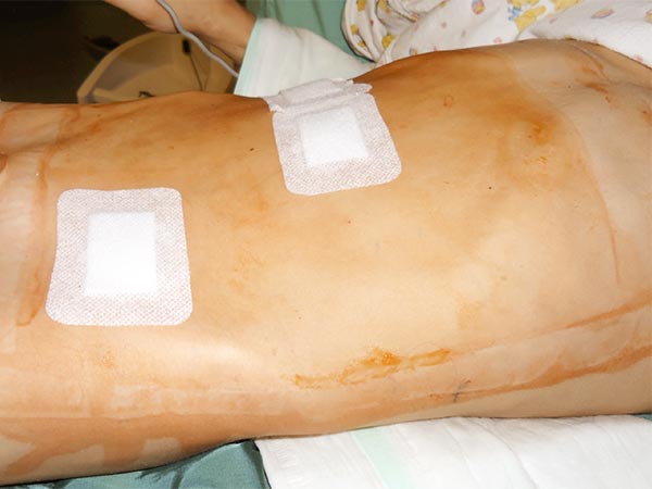
Flat abdomen at the end of surgery, no recurrence to date.
Intraabdominal, retroperitoneal, macrocystic lymphatic malformations can often be removed primarily by surgery, either completely or extensively, using minimally invasive laparoscopic surgery. Both the cases shown have been recurrence-free to date.
Published: 2018
All images © Meyer
