Chapter: Wounds and ulcerations
Article: 1 of 7
Update: Mar 10, 2021
Author(s): Ott, Hagen
Wounds are defined as skin defects which result from a separation of tissue cohesion on external or internal body surfaces and occur with or without tissue loss.
On the skin, superficial wounds located in the epidermis are called erosions, while tissue defects in the upper dermis are called excoriations. They are only referred to as ulcerations when at least the deeper layers of the dermis or underlying structures are affected.
Wound healing of external wounds means the closure of a skin defect by reconnection of the previously separated tissues. It can be roughly divided into three phases:
The term “primary wound healing” refers to almost direct, often surgical closure of wound openings, in which the above-mentioned wound healing phases occur to a lesser extent and are not very visible externally.
In “secondary wound healing”, conversely, after an initial inflammatory reaction, open skin wounds are initially bridged by granulation tissue, which is clearly visible. Not until a second step can the “repair” of the wound be seen from the renewed formation of epidermis (re-epithelialization) and possibly scar formation.
Some authors basically distinguish between three types of wounds: traumatic, iatrogenic and chronic wounds. While acute, traumatic wounds do not occur with any particular frequency in patients with vascular anomalies, both iatrogenic and chronic wounds are likely to be more common in this patient group than in the normal population.
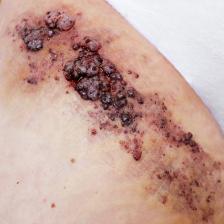
Iatrogenic wounds are caused by the physician during the course of diagnostic or therapeutic procedures. Examples include skin defects after puncture and sclerotherapy, surgery, or laser therapy. They are regrettably unavoidable in some patients receiving treatment for vascular anomalies, but they heal very often without complications under medical or nursing follow-up.
Chronic wounds persist for longer than 8 to 12 weeks and require particularly intensive wound management. Furthermore, in the case of chronic wounds, it is necessary to identify and eliminate potentially negative influencing factors (selection):
Postoperative wounds can also show a protracted healing process in patients with vascular anomalies, depending on their type, size and localization. In the case of lymphatic malformations, for example, there is a risk of recurrent accumulation and oozing of lymphatic fluid at the wound despite postoperative drain placement, which not infrequently necessitates the insertion of a second drain.