Author(s): Wohlgemuth, Walter A.
Author(s): Wohlgemuth, Walter A.
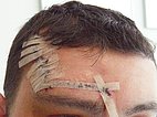
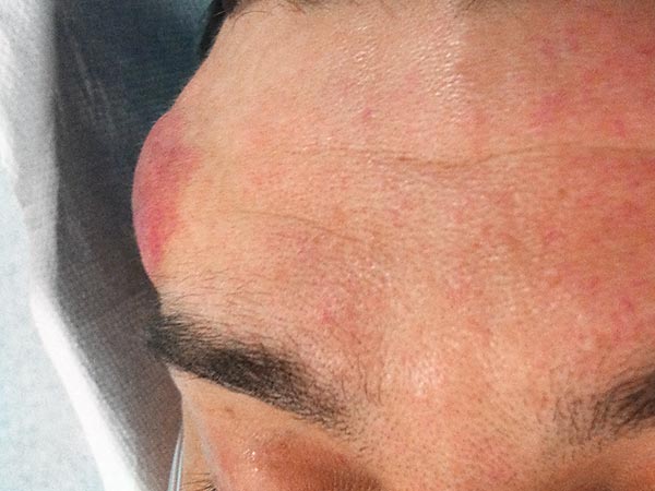
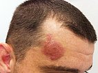
28-year-old patient with a circumscribed swelling on the right side of the forehead which had increased in volume over the past year and had previously presented as redness for a long time. The immediate surrounding skin shows a pale rim due to a steal phenomenon. On palpation, the mass is clearly hyperthermic, and a pulsation is palpable. Thus, a fast-flow malformation is likely. The photograph shows the direct top view.
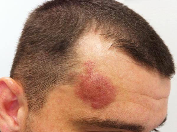
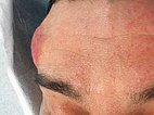
Photograph in lateral view documenting the swelling.
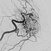
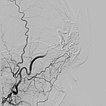
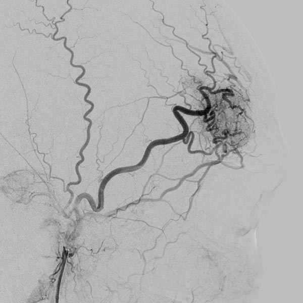
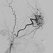
Digital subtraction angiography (DSA) with contrast injection into the right external carotid artery. The superficial temporal artery reveals a microfistulous AVM with markedly dilated feeder arteries on the right forehead with immediate venous outflow.
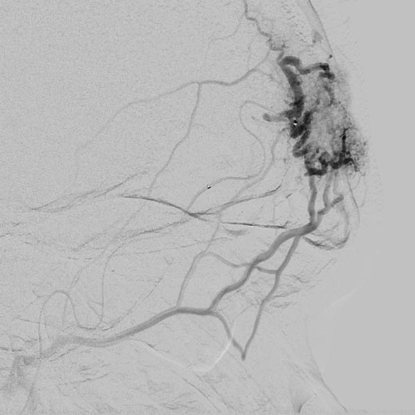
The microcatheter was advanced directly into the nidus. After visualization of the nidus, immediate direct venous outflow (DSA, venous phase) from the lesion via dilated veins. This confirms the diagnosis of an AVM.
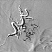
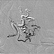
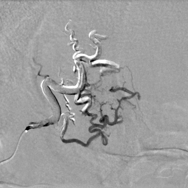
After super-selective catheterization via a microcatheter, stepwise filling of the entire nidus of the AVM using ethylene-vinyl alcohol copolymer with plug-and-push technique, in which the catheter tip is glued in place and the embolic agent is pressed into the lesion. Image in roadmap technique shows previously injected embolic agent in white and newly injected embolic agent in black. Note the increasing retrograde filling of the small feeder artery at the bottom of the lesion (start of injection).
![[Translate to Arteriovenous malformation on the forehead:] Arteriovenöse Malformation an der Stirn DSA: embolic agent is pressed into the lesion](/fileadmin/images/patientenbeispiele/5-avm-stirn/f5-05-dsa-arteriovenoese-malformation.jpg)
Note the increasing retrograde filling of the small feeder artery at the inferior border of the lesion (during injection).
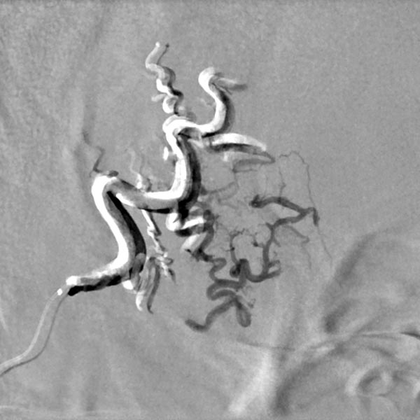
Note the increasing retrograde filling of the small feeder artery at the bottom of the lesion (after injection). The small artery was retrogradely filled against its flow direction.
DSA image depicting the arterial inflow, nidus with small arteriovenous fistulae, and venous outflow of the AVM before embolization. The complete angiomorphology of the AVM, at this point untreated, is easily visible.
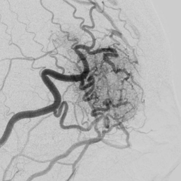
The conventional radiograph (same section as previous image) after completion of the embolization shows the complete cast specimen of the entire nidus with the radiopaque embolization material (cast). This accurately traces the anatomy and angiomorphology of the complete AVM, which is thus occluded.
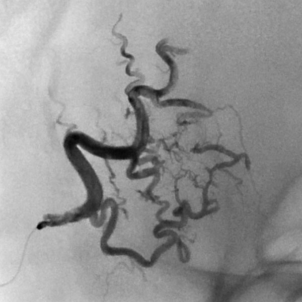
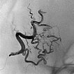
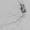
Completion angiography with contrast injection into the right external carotid artery demonstrates complete occlusion of the nidus of the arteriovenous malformation. On account of the subtraction imaging technique, the cast appears white here (subtracted).
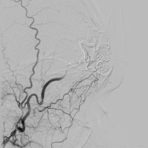
To ensure long-term success and avoid recurrence, as well as to improve the cosmetic result (persistent redness of the skin on the forehead), the complete occluded nidus was resected. To date, the patient has had no recurrence.
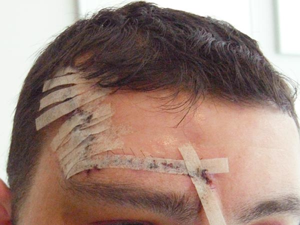
The arteriovenous malformation (AVM) on the forehead treated here, although not very large, has entered a proliferation stage (Schobinger grade II) and has shown significant enlargement in the past months. This suggests a poor long-term prognosis without treatment. In addition, there is cosmetic impairment on the forehead.
The therapeutic approach here is a combination of complete embolization and subsequent resection of the occluded nidus of the AVM.
Comparison of the initial angiography images and the image of the cast specimen of the AVM completely filled with embolic agent demonstrate the completeness of the occlusion.
In this case, the probability of recurrence is very low in the long-term course, but follow-up is still necessary in the long term.
Published: 2018
All images © Wohlgemuth
![[Translate to Arteriovenous malformation on the forehead:] Arteriovenöse Malformation an der Stirn DSA: embolic agent is pressed into the lesion](/fileadmin/_processed_/e/9/csm_f5-05-dsa-arteriovenoese-malformation_18e0bfc8c8.jpg)
