Author(s): Wohlgemuth, Walter A.
Author(s): Wohlgemuth, Walter A.
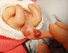
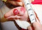
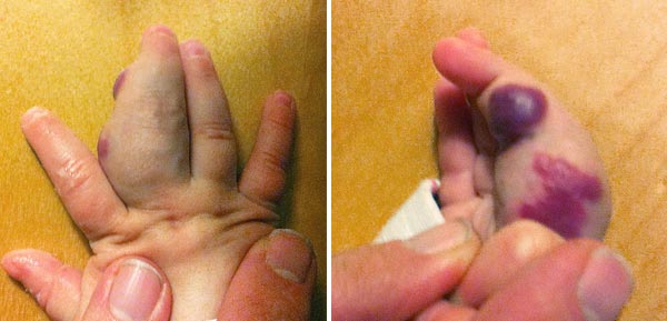
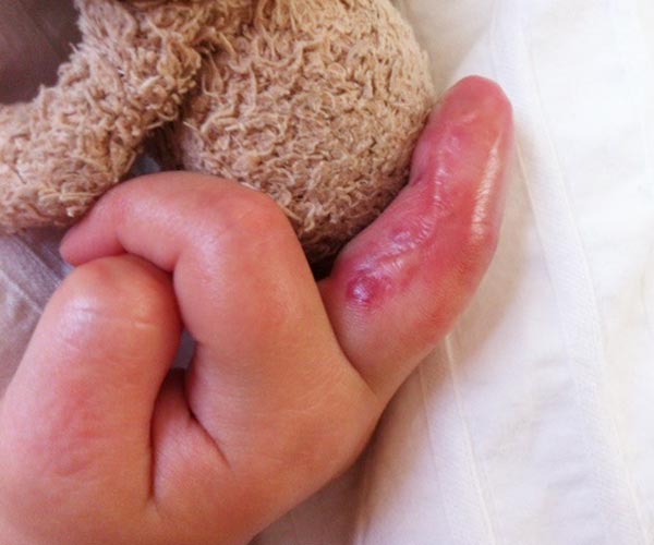
6-month-old girl with a progressive, red, pulsatile tumor on the left middle finger. The tumor is hyperthermic, thus suggesting hemodynamically relevant arteriovenous fistulas. The reddish skin discoloration initially suggested an infantile hemangioma as differential diagnosis.
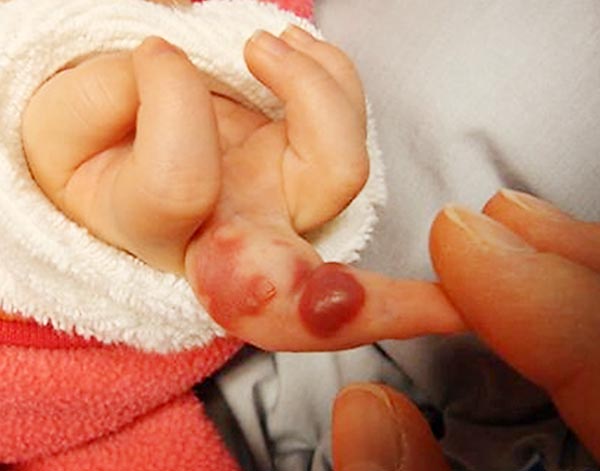
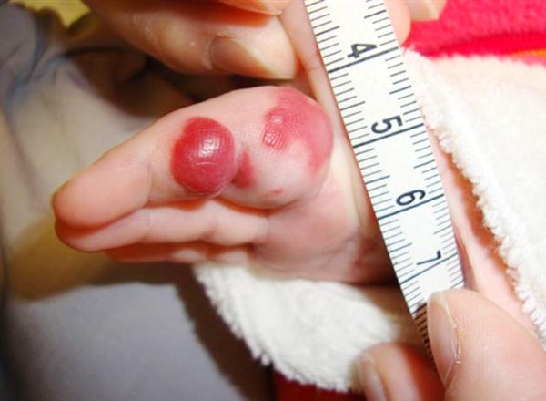
The mass also extends subcutaneously to the volar portion of the proximal phalanx and is pulsatile but relatively soft on palpation. The overlying skin is red and overheated.
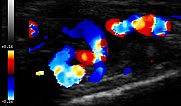
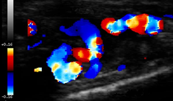
Color-coded duplex ultrasonography shows a massive increased blood flow, but not in multiple, very small reticular vessels as expected in an infantile hemangioma. The vessels were larger. This finding is more consistent with an arteriovenous malformation.
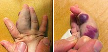
Within 4 months, there was a further increase in the size of the mass. An increasing deviation of the D3 to the lateral side with a V-shaped malposition of the D3 in regard to the D2 has developed. This added to the indication for an invasive procedure, since the full functionality of the hand is at risk.
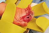
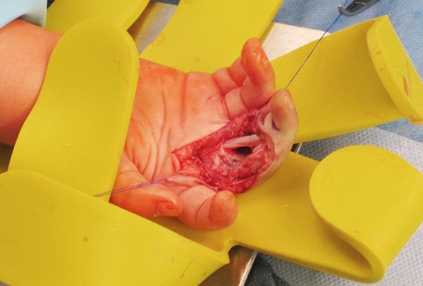
After prior percutaneous embolization, the mass was microsurgically dissected and completely removed.
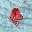
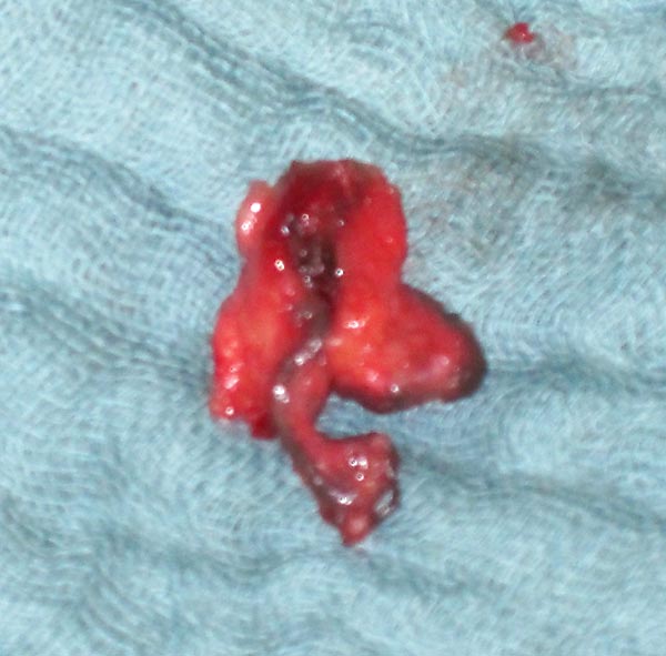
The resectate shows relatively large, dilated vessels with a relatively thick wall and the embolic agent (black intravascular material). Histologically, the diagnosis of an arteriovenous malformation was confirmed.
On the 2nd postoperative day, despite anticoagulation, peripheral ischemia occurred at the distal end phalanx of the D3 with acral necrosis; this had to be resected. The tip of the distal phalanx could not be preserved.
At clinical follow-up after 2 years and 6 months, no recurrence of AVM. The finger is fully functional, with slight lateral deviation in the proximal interphalangeal joint. However, the nail of the distal phalanx has not regrown.
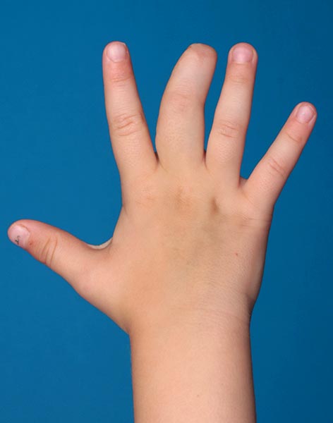
Even after 4 years and 7 months postoperatively, there is still no recurrence of the arteriovenous malformation (AVM). The middle finger has regained function and is not growing disproportionately as compared to the other fingers. This disproportionate growth would otherwise have been expected if the AVM had not been removed.

Arteriovenous malformations (AVM) rarely become symptomatic in early infancy. The rapid growth and the mass effect forced intervention in this case. The differential diagnosis from infantile hemangioma, which is also raspberry red, results from the imaging (large vessels with massive flow) and the absence of a solid vascular tumor component. Resection was greatly facilitated or made possible by prior complete embolization of the lesion. Embolization and resection, which are necessary to avoid recurrence and have to be as complete as possible, are technically difficult. In this situation it may be anatomically necessary to compromise the very distal blood supply. The initially still present blood supply of the acral fingertip was lost in the course, but the AVM could be completely removed without recurrence. In the natural course, due to continuous progression of the AVM expanding to the whole finger, the finger would have been at risk in the long run. The overall result is encouraging in view of the initially extensive AVM in this difficult localization.
Published: 2018
All images © Wohlgemuth
