Chapter: Orthopedic problems associated with vascular malformations
Article: 7 of 9
Update: Feb 24, 2021
Author(s): Kertai, Michael Amir
Various types of foot deformities occur in the context of vascular malformations.
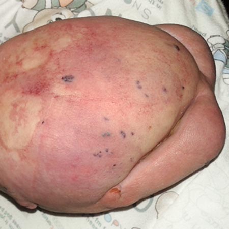
If the vascular anomaly is located directly in the foot area, the anomaly itself, depending on its extension, will cause a change in the external shape of the foot. This usually has little effect on the bony structures.
However, venous malformations in particular can lead to severe, sometimes chronic pain due to the formation of recurrent thrombophlebitis. This results in prolonged relieving of the foot by affected patients, leading to secondary contractures and atrophies of immobility. In childhood and adolescence, growth can also be slowed as a result of the low load on the extremity, which leads to a shortening of the foot or leg.
Interventional or surgical therapy of the malformation, as well as pain therapy, is the primary treatment for the problem described. In the case of localized vascular anomalies, conservative orthopedic methods such as insoles and splints (orthoses) can be used to change the pressure distribution on the foot in such a way that pain can be reduced.
Ultimately in very difficult cases, if all of the above options fail, amputation or partial amputation of the foot should be considered. In this case, a usable foot prosthesis is still better than the patient's own foot, which has no function.
Especially in the context of CLOVES syndrome, but also in other circumscribed overgrowth syndromes, sometimes pronounced deformities of the foot can occur, particularly so-called “ballooned hyperplasia” of the feet : balloon-like enlargement due to fatty tissue hyperplasias, often in combination with ray hyperplasias of individual toe rays. Massive elongations of individual toes dominate, which can be attributed to an increase in size of the metatarsal bones as well as the phalanges. In some cases, the development of an adult foot length already occurs at kindergarten age. It is not uncommon for only one part of the foot to be affected asymmetrically, the extent often differs and is unequal between the two feet.
In simpler cases, this means that only one or a few foot rays are affected. In more complex cases, there may be a juxtaposition of enlarged, normal-sized and sometimes shorter bones on one or more rays.
Often, foot enlargement is also accompanied by leg length discrepancy, as the long bones of the lower and/or upper leg are also partially affected.
If a whole ray is affected (or several), it can be removed by a ray resection. This means removing the affected metatarsal bone and the associated phalanges. This results in the formation of a four-ray foot when one ray is resected. A four- or three-ray foot still provides adequate stability, so this procedure can be performed without sacrificing function. However, this operation is distinctly limited since resection of the first and fifth rays should not be performed, as it would seriously reduce the stability of the foot.
However, if the whole foot is enlarged or only some phalanges, an individual approach must be chosen after the affected bones have been identified by radiograph.
As the growth of the tarsal bones cannot be influenced surgically, only the metatarsals and the phalanges can be addressed in a growth-restrictive operation.
Drilling out the growth plate (permanent epiphysiodesis) will stop the treated bone in its growth. It is therefore necessary to estimate the further growth of the foot when planning the operation and to perform the operation at an early enough stage in order to aim for a final foot size that can be fitted with normal shoes.
Experience has shown that destruction of the growth plate by drilling is not always successful, despite the use of correct technique. This is due, on the one hand, to the enormous growth potential of the affected bones and, on the other hand, to the fact that it can be technically difficult to identify the growth plate in the foot, which is often enlarged with soft tissue overgrowth. It is therefore advisable to check the closure of the growth plate by X-ray after one year at the latest.
As an alternative to growth retardation by destruction of the growth plate, individual phalanges can also be removed if they are locally related to the enlargement of the foot. However, in these cases, deformities of the toes often occur, so that arthrodesis of the remaining phalanges should be considered.
As a last resort, amputation or partial amputation of the foot may also be considered.
In addition, depending on the foot deformity, other interventions such as tendon transfers are possible.