Chapter: Coagulation disorders
Article: 4 of 6
Update: Feb 24, 2021
Author(s): Wohlgemuth, Walter A.
Kasabach-Merritt phenomenon (KMP) occurs almost exclusively in association with kaposiform hemangioendothelioma (KHE) or, much less frequently, tufted angioma (TA), which is histologically very similar but usually smaller and confined to the skin. Both vascular tumors (KHE and TA) are considered by many authors as one tumor entity. Previous reports of the occurrence of a Kasabach-Merritt phenomenon associated with “hemangiomas” were based on an incorrect diagnostic classification of the vascular anomaly. There are isolated reports of KMP in large angiosarcomas.
A very rare special case is the newly described multifocal lymphangioendotheliomatosis with thrombocytopenia (MLT), also called cutaneovisceral angiomatosis with thrombocytopenia (CAT) by another working group. It manifests as disseminated, usually very small, sometimes exophytic reddish vascular tumors (with lymphatic vessel endothelium) on the skin and always simultaneously in the gastrointestinal tract. MLT/CAT can also lead to severe thrombocytopenia.
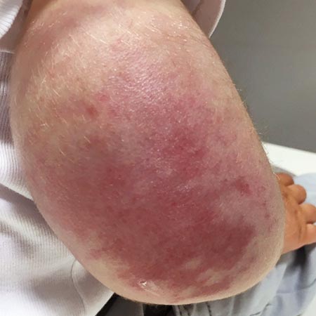
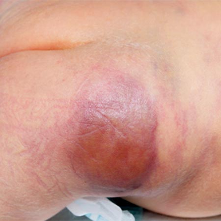
Clinically, in Kasabach-Merritt phenomenon, the vascular tumor appears indurated, reddened and shows an increase in volume, clinically similar to acute inflammation and simultaneous hemorrhage into the KHE (intratumoral coagulation and inflammation).
In severe Kasabach-Merritt phenomenon, petechiae and spontaneous hemorrhages on skin and mucous membranes additionally occur as a result of the systemic coagulation failure.
Laboratory chemistry of Kasabach-Merritt phenomenon is characterized by severe thrombocytopenia (< 20 x 109/l) with concomitant hypofibrinogenemia (< 100 mg/dl), increased fibrin degradation products (especially D-dimers), and the appearance of fragmented red blood cells (schizocytes; fragmentation by fibrin filaments).
The pathological activation of platelets in the vascular tumor and secondary activation of coagulation probably occurs as a result of the pathologically altered, dysplastic vascular endothelium within the kaposiform hemangioendothelioma. The pathological endothelium leads to platelet adhesion with subsequent platelet activation and aggregation. The half-life of a platelet is thus drastically shortened to 1 h−24 h, and platelets are consumed faster than the body can reproduce and replace them.
Secondarily, the activated platelets additionally activate plasmatic coagulation factors (especially factors V, VIII, XI), which leads to a further deterioration of the coagulation situation, since these activate fibrin formation (factor Ia) via factor IIa.
The fibrin is simultaneously degraded again via the subsequently activated fibrinolysis, and the resulting fibrin fragments cause fragmentation of erythrocytes and thus the formation of schizocytes. However, this effect on the erythrocytes is usually not so strong that anemia develops at the same time; this only happens as a result of the often massive bleeding that occurs.
If coagulation and fibrinolysis are massively activated simultaneously, full-blown disseminated intravascular coagulation (DIC) may occur. Young infants or toddlers with congenital or early-onset DIC also have an immature liver and thus a reduced synthesis capacity for replacing coagulation factors.
Since this coagulation activation with simultaneous hyperfibrinolysis and complement activation occurs predominantly in the vascular tumor, the tumor rapidly increases in size, appears plumper and warmer, as if inflamed. This effect is further enhanced immediately after transfusion of platelet concentrates. Therefore, these are only sparingly infused in cases of severe, clinically manifest thrombocytopenia, as they are also very quickly consumed again.
In more than 50% of patients with kaposiform hemangioendothelioma (KHE) or tufted angioma (TA), Kasabach-Merritt phenomenon (KMP) was described within the first year of life. In the largest cohort of 107 KHE patients, KMP was reported in a total of 71% of patients. Thus, overall, there is a relatively high probability of the occurrence of this dangerous coagulation complication in the presence of a kaposiform hemangioendothelioma.
The following are the main risk factors for the occurrence of KMP in KHE/TA:
After biopsies of complicated tumors with Kasabach-Merritt phenomenon, the histologic appearance was found to be more similar to a kaposiform hemangioendothelioma than to tufted angioma. Again, this tends to suggest that a tufted angioma may be only a less distinct subtype of a kaposiform hemangioendothelioma.
First of all, it is important to recognize the relevant risk of the occurrence of a Kasabach-Merritt phenomenon and thus establish the correct diagnostic classification of a vascular tumor as KHE/TA and not classify it as “hemangioma”.
The most important therapeutic goal is then a rapid and effective reduction of the tumor volume.
The contact area of circulating platelets with the coagulation- and platelet-activating pathological tumor vascular endothelium must be reduced as effectively as possible.
Drug therapy, radiological interventional and, in some cases, open surgical procedures are available for this purpose.
The classic combination drug therapy for KHE/TA consisted of vincristine i.v. and oral prednisolone or i.v. methylprednisolone. The length of therapy depended on tumor response, which was highly variable.
Heparin is indicated early if signs of clotting factor consumption appear.
The mTOR inhibitor sirolimus is now frequently used as primary therapy in individual cases with good to very good success, but the relevant results from prospective placebo-controlled trials are still pending. In many centers, sirolimus has already replaced vincristine completely.
Single (acetylsalicylic acid) or dual (ASA + clopidogrel) platelet aggregation inhibition is recommended in order to decrease platelet activation and aggregation. However, this does not seem to effectively inhibit a Kasabach-Merritt phenomenon.
Propranolol and interferon alpha are no longer recommended.
Antifibrinolytic drugs (such as epsilon-aminocaproic acid or tranexamic acid) have also shown little efficacy in the published literature with regard to bleeding in Kasabach-Merritt phenomenon.
Caution: Transfusion of platelets enhances trapping of platelets in the tumor and leads to a rapid increase in tumor volume, with signs of inflammation and increasing pain, as well as increased coagulation activation and consumption. The half-life of platelets transfused in this process is extremely short, and massive local platelet activation results in additional local release of proangiogenic factors with potential tumor enlargement. Therefore, the use of platelet concentrates should be strictly limited.
In cases of manifest hypofibrinogenemia (< 100 mg/dl), administration of fibrinogen or fresh frozen plasma (FFP) is indicated with concomitant administration of heparin. This is especially true in connection with clinically manifest bleeding.
For rapid improvement of the coagulation situation, invasive therapeutic measures may be justified in infants and young children.
Open surgical resection of the vascular tumor is usually not possible or only partially possible in the acute stage. This is due to the often diffuse infiltration of the kaposiform hemangioendothelioma through several tissue layers, the poor demarcation from non-infiltrated tissue, the concomitant presence of lymphedema, and the sometimes large extent of the tumors at an anatomically unfavorable site. This is further hampered by the co-existence of a very difficult coagulation situation and the fact that the overall condition of the patient in coagulation failure is often unstable.
Interventional radiological, percutaneous, transarterial embolization therapy of KHE/TA with the goal of at least partial devascularization represents a good invasive alternative, especially in the clinically difficult coagulation situation of a Kasabach-Merritt phenomenon. Interventional devascularization leads directly to significantly lower platelet activation. This is due to reduced contact of the platelets with the pathological tumor vessel endothelium; in some cases, depending on the radicality of the embolization, this platelet contact may even be completely absent. Coagulation parameters can improve massively as early as 24 hours after embolization. This embolization is usually performed as particle embolization via microcatheter under prophylactic anticoagulation. Special knowledge in pediatric interventional radiology is helpful here; the procedure is reserved for appropriately experienced interdisciplinary centers with cooperation between interventional radiology, pediatrics, pediatric surgery, and anesthesiology.