Author(s): Wohlgemuth, Walter A.
Author(s): Wohlgemuth, Walter A.
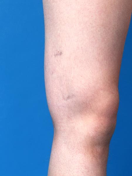
12-year-old female patient with circumscribed, relatively soft palpable swelling on the lateral distal thigh. Dysplastic veins are visible on the skin in 2 places. In the area of the swelling, recurrent induration and circumscribed pain due to thrombophlebitis in the venous malformation.
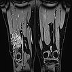
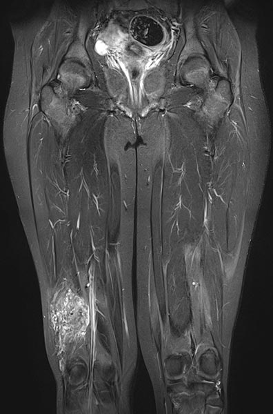
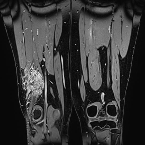
Coronal fat-suppressed T2-weighted MRI of the thighs shows an intramuscular venous malformation on the right thigh (hyperintense).
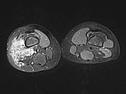
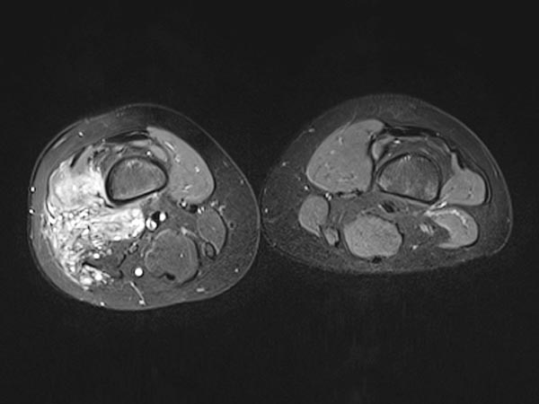
Axial T2-weighted MRI, fat-suppressed, shows the close relationship to the periosteum of the femur. This location is particularly painful due to inflammatory irritation of the periosteum.
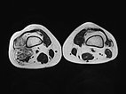
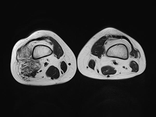
T2-weighted MRI without fat suppression in the axial plane reveals inflammatory, fibrotic remodeling of the lesion due to multiple inflammations. The right vastus lateralis of the quadriceps femoris muscle is completely penetrated by the lesion.
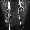
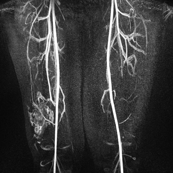
Dynamic MR angiography 62 s after injection of the contrast agent shows slow pooling of the contrast agent in the lesion without early venous return (slow-flow malformation). The lesion is connected to the deep conducting vein system (communicating veins).
The VM completely enhances contrast media (MRI, T1-weighted, fat-saturated). This makes the differential diagnostic consideration of a lymphatic malformation redundant.
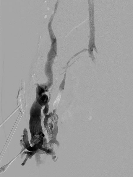
Digital subtraction angiography after direct puncture of the venous malformation during sclerotherapy. In addition to the lesion, direct communicating veins with the deep conducting vein system are visible. These were occluded with viscous alcohol gel by direct puncture.
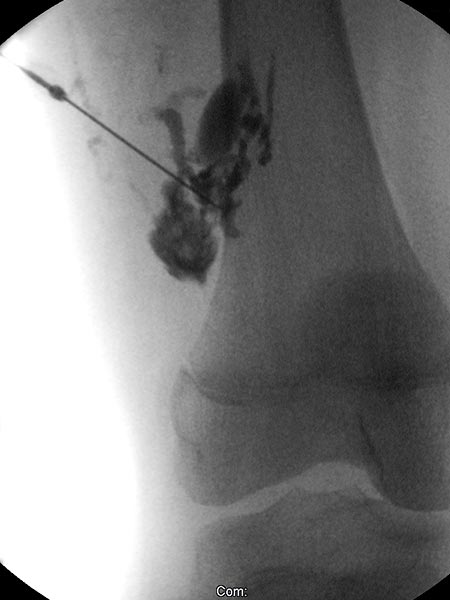
X-ray fluoroscopy after direct puncture of the venous malformation during sclerotherapy. As a result of closure of the communicating veins to the deep venous system, the VM is now isolated and can be sclerosed. After injection of contrast, no outflow of the contrast medium into the deep veins.
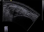
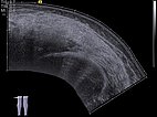
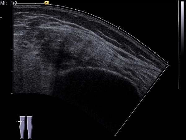
Ultrasound image (automatically assembled 2D image) before sclerotherapy. The cavities of the venous malformation are initially hypoechoic to echo-free. These cavities will be occluded by the inflammation induced by the sclerotherapy.
Ultrasound image (automatically assembled 2D image) after sclerotherapy. The hypoechoic cavities of the venous malformation are now occluded by the inflammation induced by sclerotherapy. The image now appears more homogeneous and echogenic on ultrasound. After occlusion of the cavities, painful thrombophlebitis can no longer occur.
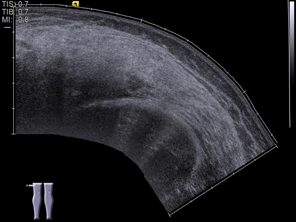
Intramuscular venous malformations (VM) are often located in the distal thigh, near the periosteum of the femur. Symptomatology of recurrent pain with accompanying induration leads to imaging, and MRI confirms the diagnosis. During sclerotherapy, it is important to exclude draining or communicating veins of the venous malformation into the deep conducting venous system, as thromboembolic processes can occur via the communicating veins either spontaneously or induced by the therapy. Sclerotherapy occludes the initially open cavities of the VM, which thus becomes more echogenic in ultrasound. Today, the patient is asymptomatic even 2 years and 4 months after the therapy.
Published: 2018
All images © Wohlgemuth


