Author(s): Cangir, Özlem
Author(s): Cangir, Özlem
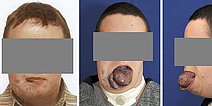
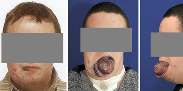
18-year-old patient with bluish spongy, enlarged tongue and lower lip on the right side and palpable thrombophleboliths in the area of the right cheek due to an extensive venous malformation (VM). A venous malformation can also be seen on the skin of the right cheek.
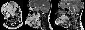
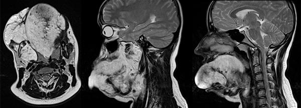
Sagittal and axial T2-weighted MRI of the face shows the extensive venous malformation located in the whole tongue and right cheek muscles.
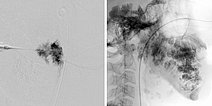
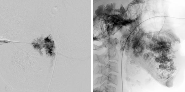
Digital subtraction angiography (DSA left; unsubtracted image right) after direct puncture and insertion of a cannula into the venous malformation during sclerotherapy.
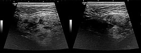
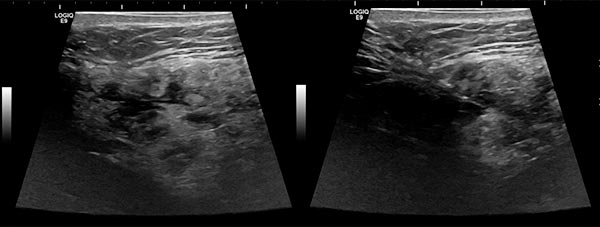
Ultrasound images (automatically assembled 2D B-mode images) before sclerotherapy. The initially hypoechoic cavities of the venous malformation are occluded by the inflammation induced by sclerotherapy.
Outcome after sclerotherapy, tongue wedge resection, laser therapy, and lip correction surgery as well as repeat sclerotherapy treatments after a total of 7 procedures (including tracheostoma creation and closure) and 10 months’ total treatment duration.
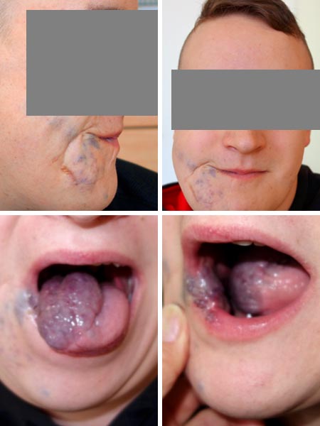
Venous malformations (VM) located in the tongue and cheek/lip region may extend into the pharynx. A safe upper airway is required for treatment, for which a tracheostomy is sometimes required if there are extensive findings. Furthermore, implantation of a central venous catheter and prolonged postoperative or post-interventional intensive care may be required.
An extensive venous malformation may result in localized intravascular coagulation (LIC) with elevated D-dimers and low fibrinogen. This requires close hemostaseology/pediatric monitoring and therapy.
If left untreated, the venous malformation will lead to continued enlargement of the affected veins and complicate therapeutic access.
Published: 2018
All images © Cangir
