Chapter: Venous malformations
Article: 3 of 13
Update: Mar 25, 2021
Author(s): Barbera, Letterio Christian
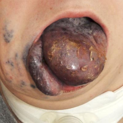
A thorough clinical history and a thorough physical examination are indispensable for determining the respective characteristics of an individual venous malformation. Information is sought about when the abnormalities first appeared clinically and how they have changed over the years or as a consequence of previous treatments. Bluish discoloration of the skin, prominent visible vessels and circumscribed swelling can occur singularly or together. As parents are usually very familiar with their children’s bodies, they should be given time to report. In addition to appearance, function and pain play an important role in understanding the very individual clinical picture. For example, parents should be asked about everyday activities such as playing, running, writing. Does the child avoid certain movements or does he or she behave like their peers at school or during sports? Specific questions are asked about complaints in the affected part of the body. The duration, frequency, type and intensity of the pain should be determined and documented. Sudden onset of localized and severe pain lasting a few days indicates thrombophlebitis in the venous malformation. A recurrent and dull, tight pain in combination with a feeling of pressure indicates congestion of the immature vascular areas. Frequency and intensity of the pain (on a scale of 1–10) should be documented. This is important in deciding whether invasive treatment is indicated.
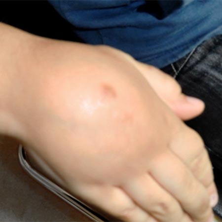
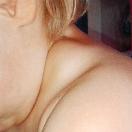
The physical examination begins with an inspection of the skin, which often reveals the extension of the venous malformation. Irregularly shaped, dilated and bluish vessels are visible on or shimmer through the skin. Their diameter is 1–30 mm and depends on body position. They fill up with blood when the limb is suspended. A venous malformation on the forehead, for example, becomes particularly prominent when the head is bent over. These vessels are usually soft, compressible and not painful. The findings are different in thrombophlebitis: In this case palpation is painful and the vessel is indurated. A capillary malformation (port wine stain) is frequently observed, in which case it is a combined capillary-venous malformation (CVM). The spread of the port wine stain usually coincides with expansion of the venous malformation in deeper tissue layers.
Attention should also be paid to posture and movement. More deeply located venous malformations near a joint cause pain during movement and force the sufferer to adopt a relieving posture of the affected extremity. There is pain in the soft tissues or joints of the leg, which the patient will often elevate spontaneously. Venous malformations in the subcutis are rarely the cause of complaints related to impaired movement and physical strain. In these cases, subfascially located, intramuscular venous malformations are responsible for pain and functional impairment. When an affected limb is elevated, the volume of the affected limb often decreases substantially thanks to blood outflow.
It is always important to compare the length and circumference with the opposite side. Different circumferences can be caused by a voluminous venous malformation, by an additional lymphatic malformation, but also by partial hypoplasia or overgrowth of the soft tissue. If the circumference of an arm or leg decreases with elevation, an extensive venous malformation is probably present subcutaneously and subfascially. An accurate clinical examination of the subcutis and the underlying musculature can usually determine the cause of the difference in circumference.
If increased warmth and a thrill are detected on palpation of an affected area, this does not indicate a venous malformation, but an arteriovenous malformation (AVM) as a fast-flow malformation. In this case, the visible, prominent veins are not immature, dysplastic veins but regular veins that have become hyperplastic because of increased blood flow and blood pressure (arteriovenous shunts). In contrast to venous malformations, the vascular wall is thickened and the shape is more regular and tubular. The attending physician determines which changes may be responsible for the symptoms. The unique, individual characteristics of a venous malformation in each patient should first be assessed on the basis of the clinical presentation. Only then can the appropriate diagnostic modality and therapy be planned and initiated.