Chapter: Conservative therapy
Article: 2 of 8
Update: Mai 15, 2021
Author(s): Wohlgemuth, Walter A.
It can sometimes be difficult without imaging to make a clinical diagnosis of edema (whether lymphatic or venous) versus circumscribed soft tissue proliferation (mostly fat) in patients with vascular anomalies and enlarged extremities. This is especially true when a syndromic vascular malformation with additional soft tissue proliferation is present. The distinction is important, however, because compression therapy is much more effective in edema than in circumscribed tissue hyperplasia.
In principle, the clinical signs in the patient history of edema and thus the increase in volume tends to fluctuate, sometimes more and sometimes less. Pure soft tissue proliferation (mainly caused by circumscribed fat tissue proliferation) is rather static and changes only very slowly (increase), if at all. Additional imaging is very helpful in the differential diagnosis, especially sonography and MRI which can reliably distinguish between the two changes (soft tissue hyperplasia versus edema). Lymphoscintigraphy with injection of Tc99 colloids (albumin or sulfur) into the interdigital spaces between the toes is also helpful in special cases and can reliably detect a pathological slowing of lymphatic flow in the leg. In pathological cases, the colloid arrives in the inguinal lymph nodes on the affected side only after more than 1 hour, thereby confirming the diagnosis. Direct intranodal lymphangiography is a new, very promising diagnostic tool for changes in the central lymphatic conductors of the trunk of the body.
Lymphedema in lymphatic malformations differs from phlebedema in venous malformations, among other things, by the additional deposition of proteins in the superficial interstitial tissue spaces. If left untreated, lymphedema in the late stage leads to an increase and hardening of the superficial tissue and functional disorders of the skin through fibrosis and the storage of additional tissue in the extracellular space.
Primary lymphedema in the sense of a congenital lymph transport disorder in people under 20 years of age affects about 1.2 out of 100,000 people. However, significantly more than 99% of all lymphedema worldwide is secondary, e.g., after infections (filariasis) or following operations or radiation, etc.
Both types of edema (lymphedema and phlebedema), however, respond positively to compression therapy. Intensive compression also has a positive effect on tissue hyperplasia, although it is less effective than in edema.
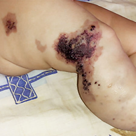
Blood pressure measurements and venous punctures may only be performed on a lymphedematous limb if a healthy limb is no longer available, as long as this is done under sterile conditions and extravasation is avoided.
A basic distinction is made between static compression therapy, which is always applied in the same way, and dynamic compression therapy, for example, with massages or pneumatic technical compression pumps, which also achieve a dynamic effect. The two procedures are often combined: first edema mobilization by dynamic procedures, then obtaining the result by static bandages or compression garments.
The aim of compression therapy is to permanently reduce fluid in the tissue volume, usually of a limb, and to reduce of progression of the edema, particularly preventing lymphedema from developing into the irreversible form with irreversible hardening of the skin and subcutaneous tissue owing to the deposition of proteins.
Compression therapy performed over many years can also be used in cases of soft tissue hyperplasia with fatty tissue hyperplasia. Strict monitoring, especially of the growing limb, is required.