Chapter: Vascular tumors
Article: 2 of 11
Update: June 02, 2021
Author(s): Evert, Katja | Ott, Hagen
Infantile hemangiomas (IH) are very common vascular tumors that initially grow and later regress spontaneously following a typical phase sequence. Their antigen structure is similar to that of placental tissue. Local or regional tissue hypoxia is discussed as a possible pathogenic factor in their development. In most cases they regress spontaneously, but changes in the skin or subcutaneous tissue can persist. They must be distinguished from other vascular tumors, e.g., kaposiform hemangioendothelioma (KHE) or pyogenic granuloma, and from arteriovenous, venous, lymphatic or combined malformations of the vascular system. In histology specimens, which are rarely required in clinical routine, one of the ways to make this distinction is by immunohistochemical detection of the glucose transporter GLUT1.
As the most common benign skin tumors in childhood, infantile hemangiomas occur in up to 5% of all infants. They are characterized by their typical growth pattern and manifest themselves only after birth. In the vast majority of cases, they become visible after a few weeks of life as enlarged, often somewhat raised, raspberry-red spots on the skin with sharp edges. They rapidly increase in size during the following weeks and months of life (proliferation phase). They then undergo growth arrest (plateau or stagnation phase), until finally around the first birthday spontaneous regression begins (regression phase), which is very often completed between the fourth and eighth year of life. However, it is not uncommon for local teleangiectasia, skin pigmentation, cutis laxa or soft, fibrolipomatous soft tissue proliferations to remain, especially in large, ulcerated or untreated infantile hemangioma. Because of this spontaneous regression, which occurs in a high percentage of affected children, invasive treatment of the infantile hemangioma is usually not necessary.
The adjacent picture (Photograph of a typical uncomplicated infantile hemangioma...) shows an uncomplicated infantile hemangioma in the area of the right wrist, which does not require specific therapy in view of the expected spontaneous regression.
This group of unproblematic hemangiomas must be distinguished from complicated forms of infantile hemangioma, where the beta-blocker propranolol is approved for the medical treatment of infants:
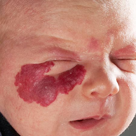
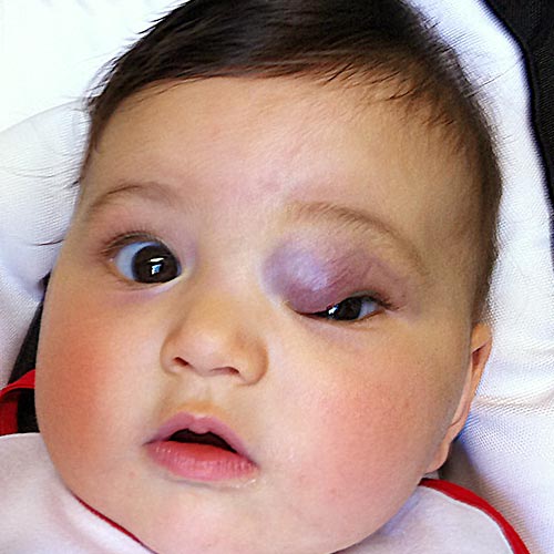
Infantile hemangiomas in an unfavorable localization are associated with a risk of functional impairment. This is particularly true for infantile hemangioma in the mid-facial region, such as on the eyelids, which can lead to visual impairment or even blindness of the affected eye.
In addition, infantile hemangioma localized on the lips can be associated with feeding difficulties during infancy. In addition, infantile hemangiomas at the tip of the nose often grow rapidly and can lead to a disfiguring, permanent deformation of the nose. Furthermore, infantile hemangioma in the above-mentioned, highly visible sites can be associated with a psychosocial burden on affected children and their parents.
Ulcerative infantile hemangiomas are associated with deeper-lying skin defects and occur preferentially on the lips and in body folds, especially in the diaper area, armpits or neck creases.
Ulcerations are often very painful, have an increased risk of infection and can heal with permanent scarring. They should therefore be treated early. Small and superficial ulcerations can often be controlled by careful wound management. If this therapeutic approach remains unsuccessful, oral treatment with propranolol should always be initiated, particularly if there is associated pain.
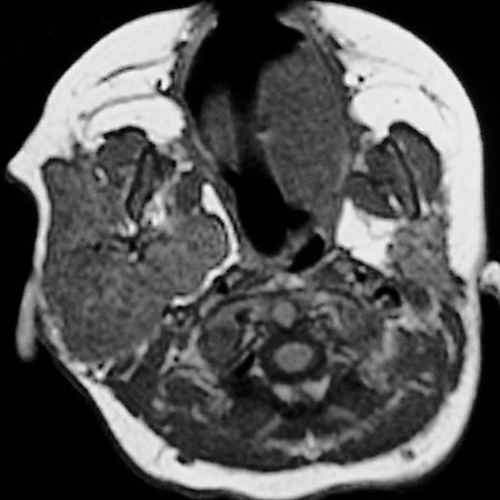
Segmental infantile hemangiomas often involve rather extensive, specific anatomical regions along embryonic, neuroectodermal units (e.g., in the face, lumbosacral). They account for about one-seventh of infantile hemangiomas and may occur in association with other malformations. The best known example of complicated, segmental infantile hemangioma is known as PHACE syndrome. The abbreviation PHACE here describes the common occurrence of brain malformations in the area of the posterior fossa with a large hemangioma in the facial region as well as eye malformations and blood vessel or heart anomalies. As with other infantile hemangiomas in the face, cosmetic and psychosocial problems arise first. In addition, individual involved segments are associated with typical risks. For example, the upper or lower eyelid can be affected and lead to visual impairment or even blindness. In addition, PHACE hemangiomas of the middle lower jaw, known as the beard region, can also be accompanied by additional hemangiomas in the trachea, making further diagnosis (laryngotracheoscopy) necessary. Infants with PHACE syndrome also require propranolol therapy, which should be initiated at an experienced center.
Multifocal infantile hemangioma, also known as neonatal hemangiomatosis, is diagnosed when an infant develops more than five, according to some experts more than ten, infantile hemangiomas. While the majority of affected children show no other abnormalities, in some cases additional infantile hemangioma may occur in internal organs. This must be diagnosed (e.g., by ultrasound). However, serious complications such as bleeding in the gastrointestinal tract or high-output cardiac failure due to numerous liver hemangiomas are fortunately rarely observed. The therapy of choice for symptomatic multifocal infantile hemangioma with organ involvement is systemic propranolol administration.