Cystic lymphatic malformations (LM) may be the main feature determining the symptomatology of a clinical picture, such as in Gorham-Stout syndrome which is characterized by progressiveosteolysis due to lymphatic vessel proliferation. The cause is as yet unknown.
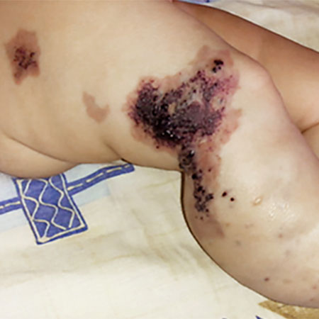
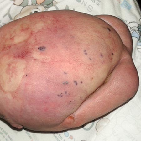
One lymphatic malformation in children that involves the abdomen is Central Conducting Lymphatic Anomaly (CCLA). This is defined as inadequate central drainage of the major trunk lymphatics due to dysmotility, stenosis, or aplasia of the main central abdominal and/or thoracic lymphatic ducts. Insufficient drainage due to congenital malformations of the thoracic duct or cisterna chyli results in lymphostasis with consecutive peripheral lymphatichypertension and reflux in various organ systems, depending on the location of the drainage obstruction. In this context, thoracic lymphatichypertension can lead to pulmonarylymphatichypertension with recurrent chylous pleural effusions or lymphaticcongestion of the lungs with plastic bronchitis. Abdominal outflow obstruction (e.g., dysplasia or aplasia of the cisterna chyli) can result in protein-losing enteropathy, chylous ascites, abdominal lymphaticcysts, and/or reflux and retention of lymphatic fluid into one or both extremities (lymphedema of the leg) or the genital region. The pent-up lymphatic pressure can in turn lead to leakage of lymphatic fluid through the skin (lymphorrhea) or cutaneouslymphatic vesicles. Intranodal lymphangiography with ethiodized poppy seed oil injection into a punctured lymph node in the inguinal region can be very helpful in diagnosing this serious condition.
Therapy: MEK inhibitor, interventional radiology. Cause: somatic ARF mutation (X-linked), which activates the MAPK pathway.
Syndromes with disorders of the lymphatic drainage system (primary lymphedema)
Syndromal forms of lymphedema include autosomal dominant as well as autosomal recessive forms.
Nonne-Milroy-Meige syndrome (hereditary lymphedema type I (Nonne-Milroy) and type II (Meige))
Lymphedema of the lower extremities, usually bilateral, may be asymmetric; occasionally confined to one foot or individual toes
Swelling initially soft, progressively hard with hyperkeratosis and papillomatosis
Upwardly curved nails
Inconsistent: large-caliber, prominent leg veins
Inconsistent: hydrocele and anomalies of the urethra in males
Cause: heterozygosity for mutation in FLT4 gene (= VEGFR3), clinically indistinguishable from mutations in VEGFC gene or GJC2 gene, autosomal dominant inheritance
Lymphedema-distichiasis syndrome
Lymphedema of the lower extremities, usually asymmetric; in males, possibly with significant scrotal swelling
Frequently venous insufficiency
Double rows of eyelashes on upper and lower eyelids in 90-95% (from the openings of the meibomian glands)
Cause: heterozygosity for FOXC2 mutations, autosomal dominant inheritance
Congenital microcephaly, decrease of 2 to 6 standard deviations
Inconsistent: congenital lymphedema, especially lower extremities (dorsum of foot)
Inconsistent: chorioretinal dysplasia with lacunar foci outside the macula, variety of other ocular anomalies
Inconsistent: mild to severe mental retardation, spasticity, epilepsy
Cause: heterozygosity for KIF11 mutations, autosomal dominant inheritance
Emberger syndrome (primary lymphedema with myelodysplasia)
Primary lymphedema (manifestation in childhood) of the lower extremities (unilateral, bilateral), often also of the genitals
Hematologic changes: myelodysplasia / acute myeloid leukemia (often with monosomy 7) and beginning as pancytopenia
Multiple warts
Hearing loss
Minor anomalies (hypotelorism, nuchal folds, slender fingers)
Cause: heterozygosity for GATA2 mutations, autosomal dominant inheritance
Noonan syndrome
Lymphedema prenatally (neck edema, chylothorax, hydrops) and/or postnatally (hand/foot dorsal edema of the newborn, lymphedema of the legs or genital area in adults)
Heart defects (60-70%)
Short stature/low normal height (-1.5 to -3.5 standard deviations)
Craniofacial anomalies
Wide thorax with pectus carinatum/excavatum
Statomotor and mild intellectual development disorder; IQ 85-90
Cause: heterozygosity for GATA2 mutations, autosomal dominant inheritance
Hennekam syndrome
Primary lymphedema (face, lower limbs, genitals).
Intestinal lymphangiectasia > exudative enteropathy (protein loss) with growth retardation, peripheral edema, and ascites
Facial dysmorphia: roundish face with flat profile, hypertelorism, blepharophimosis, wide flat nasal bridge, long flat philtrum, small mouth. Low-set small ears with thick helices