Chapter: Capillary malformations
Article: 4 of 14
Update: Mar 30, 2021
Author(s): Ott, Hagen
In order to accurately assess the clinical presentation of all patients with capillary malformation (CM), a detailed medical history and a careful clinical examination of apparently “trivial” looking skin lesions are essential. If the extremities are affected, the length and circumference of the affected extremity should be recorded in comparison to the opposite side. In addition, standardized photographic documentation is advised both in the natural history of the disease and in the case of laser therapy.
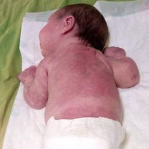
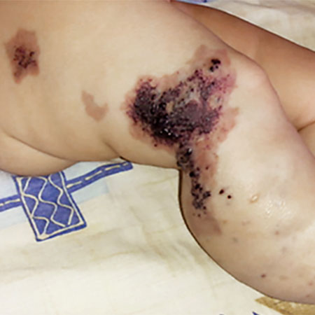
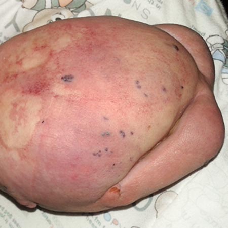
Capillary malformations cannot be visualized by prenatal ultrasound or other imaging modalities. However, they are usually visible with the naked eye immediately after birth, even though small spots in particular may not be noticeable until subsequent days of life. In affected newborns and young infants the capillary malformation appears as sharply demarcated macules ranging in color from light pink to dark red which rarely cross the midline, are often bizarrely shaped and are blanched by diascopy or finger pressure. Other manifestations of capillary malformation include reticular (net-like) patches, for example, in cutis marmorata telangiectatica congenita or MCAP. In rare cases, capillary malformations are surrounded by a lighter halo and have macroscopically visible dilated small skin vessels (teleangiectasia). Simple capillary malformations are not palpable and do not show hyperthermia compared to the surrounding skin. On auscultation no pathological findings, especially no thrill, are heard in isolated capillary malformation.
The temperature-dependent change of the color of the skin manifestations is typical of a capillary malformation. The color changes more to reddish in warm conditions (e.g., in a warm shower) and more to bluish in the cold. This temperature-dependent color change is particularly pronounced in the first years of life and then usually fades. In the natural course of development, capillary malformations may spontaneously fade and become less visible until the adolescent years, but they may also change their hue.
Capillary malformations occur most frequently in the head and neck region. In this region (e.g., the CM of Sturge-Weber syndrome) they do not show a random distribution, but follow segmental developmental patterns as cutaneous mosaics that rarely cross the midline. The traditional description of the distribution of facial capillary malformations along the three main sensory branches of the fifth cranial nerve (V1, V2, V3), the trigeminal nerve (see fig.), has been superseded, based on recent studies, by the view that facial capillary malformations can also follow circumscribed embryological developmental areas of the course of the blood vessels. Sturge-Weber syndrome (SWS) occurs more frequently in patients with a capillary malformation in the region marked with an arrow and highlighted in blue in the figure.
The corresponding skin lesions range from isolated, small, bright red spots to an extensive, dark red discoloration of an entire half of the face and the adjacent neck region.
In cases of extensive capillary malformation in the peri-orbital or peri-oral region, it is not uncommon for the adjacent mucous membranes to be affected as well.
On the trunk and extremities, capillary malformations may show configurations where postzygotic mutant “vascular cells” are distributed along embryonic developmental lines, known as Blaschko’s lines. Broad lines, leaf-like, fountain-like, or segmental patterns can develop in the process.
Other manifestations are small-spotted (e.g., CM-AVM), large segmental, sometimes “geographic” (e.g., Klippel-Trénaunay syndrome, CLOVES, Proteus syndrome), or checkerboard-like.