Chapter: Capillary malformations
Article: 7 of 14
Update: Mar 30, 2021
Author(s): Ott, Hagen
First of all, isolated cutaneous capillary malformations (CM) must be distinguished from the special types, nevus simplex, cutis marmorata telangiectatica congenita (CMTC) and teleangiectasias.
The most common and, from the therapeutic point of view, highly relevant differential diagnosis is infantile hemangioma, which can also show a segmental configuration as a variant in the cervicofacial region and on the extremities (e.g. PHACES syndrome). Classic distinguishing features of infantile hemangiomas and capillary malformations are summarized in the following table.
| Distinguishing feature | Capillary malformation | Infantile hemangioma |
| Time of manifestation | At birth | Frequently in weeks 2 to 4 of life, only rarely at birth |
| Growth | Proportional “growing” along with general body growth | Rapid growth during months 2 to 6 of life |
| Morphology | Spotty, non-elevated skin redness | Initially flat, rapidly increasing in volume, red plaques |
| Ulceration | None | Rarely, especially when occurring on the lips and in body folds |
| Responds to propranolol therapy | No | Yes |
Capillary malformations may be partial manifestations of complex clinical conditions. Despite great clinical variability, further imaging examinations should be considered in the case of the following clinical signs:
Although rare at birth and in early infancy, linear circumscribed scleroderma (morphea) should be considered as an important differential diagnosis for capillary malformations in the face and neck region of newborns. After initially appearing as a linear red patch of facial skin, the skin in morphea shows hardening and atrophy as it progresses. Thus a biopsy followed by histopathologic tissue examination should be performed early if these symptoms appear.
Cutaneous AVM is also occasionally visible as reddish, patchy discoloration of the skin. However, it is usually more indistinctly demarcated and shows clinically (hyperthermia) and sonographically (especially in color-coded duplex sonography) marked hyperperfusion, often including dilated subcutaneous vessels.
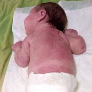
Cutis marmorata telangiectatica congenita (CMTC) has a more reticular configuration and consists of a reddish, marbled pattern with intervening normal areas of skin. It may also be combined with capillary malformation.
Nevus simplex occurs in at least one-fifth of all newborns, mostly in the nuchal region (“stork bite”), on the eyelids and mid-forehead region, or between the eyebrows. Compared to a capillary malformation, nevus simplex is light or salmon red and often has blurred margins to the surrounding skin.