Chapter: Arteriovenous malformations
Article: 2 of 13
Update: June 02, 2021
Author(s): Uller, Wibke
The specific clinical presentation of an arteriovenous malformation (AVM) can be derived from its specific hemodynamics:
The shunt as a direct, pathological connection between the arterial high-pressure system and the venous low-pressure system creates a pathological connection along the pressure gradient that bypasses the capillary bed. This shunt has significantly lower blood flow resistance than normal physiological flow with an interspersed capillary bed between them. As a result of the lower flow resistance, more blood flows via the pathological shunt, i.e., the arteriovenous fistula or arteriovenous malformation, than via the normal capillary bed in healthy tissue with its higher flow resistance.
This pathologically increased blood flow (shunt) initially leads to dilatation of the arteries leading to the arteriovenous malformation (“feeder arteries”), which may also exhibit degenerative vessel wall changes with dilatation and aneurysm formation (so-called "flow-related aneurysms") in the course of the disease.
In addition, the draining veins of the arteriovenous malformation are exposed to increased blood volume and blood pressure. Thus dilatation occurs with corresponding morphological and functional changes to the vein wall. The increased pressure load in the venous system also has a negative effect on the veins distal to the lesion.
These conditions lead to lower flow resistance in the arterial system and increased venous pressure distal to the arteriovenous malformation. This results in reduced tissue perfusion pressure with a relative undersupply of arterialized, oxygen-rich blood to the tissue and additional chronic venous insufficiency. The heart reacts to the reduced arterial flow resistance caused by the shunts of the arteriovenous malformation by increasing cardiac output. The relevant consequences are listed in the special chapter on cardiac complications.
As the individual size, angioarchitecture and localization of arteriovenous malformations differ, the course of the disease can vary significantly from patient to patient. Despite the tendency always to follow a progressive course, there can also be symptom-free intervals with an arteriovenous malformation. However, spontaneous, natural regression of this clinical picture can hardly ever be expected.
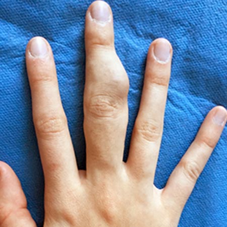
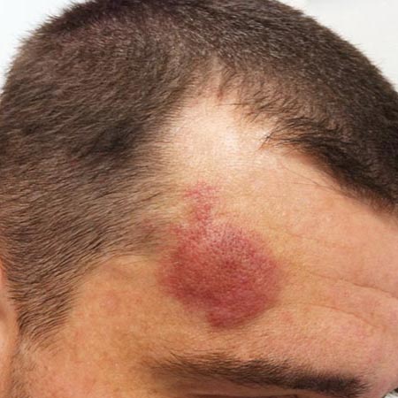
Most superficial arteriovenous malformations are diagnosed in childhood, but 8.5% of arteriovenous malformations are diagnosed in puberty. During childhood the fast-flow properties of the arteriovenous malformation may not yet be well developed. As a result, when they involve the skin in this phase of life, they are often misdiagnosed as infantile hemangiomas or capillary malformations. However, about 21% of arteriovenous malformations are still not correctly diagnosed even in adulthood.
Overall, arteriovenous malformations are very difficult to treat because of their frequent, albeit often slow, progression, their tendency to recur with incomplete therapy and the possibility that they will worsen with inadequate therapy. Among all vascular malformations they pose the greatest challenge for patients and physicians.