Chapter: Arteriovenous malformations
Article: 6 of 13
Update: June 02, 2021
Author(s): Uller, Wibke
A simple capillary malformation (CM) is characterized by a circumscribed reddish discoloration of the skin with sharp borders and without any other symptoms. It is often clinically possible to distinguish it from an arteriovenous malformation because the increased skin temperature as well as increased pulsatility and visible ectatic subcutaneous vessels typical of AVMs are absent. On imaging, capillary malformation is clearly limited to the skin level and is not usually visible at all on account of its thinness. By contrast, arteriovenous malformations can affect all tissue layers and organ systems and always have a nidus with clearly dilated inflow and outflow vessels.
In the case of traumatically acquired arteriovenous fistulas (AVFs) or aneurysms, the medical history is often indicative. A classic reticular nidus cannot be detected on imaging in the early stages. The shunt area is more circumscribed and usually related to a trauma. However, a traumatic arteriovenous fistula can progress over time into a lesion similar to an arteriovenous malformation. In these cases, a detailed medical history is indispensable, since imaging alone cannot always detect the cause of the arteriovenous malformation.
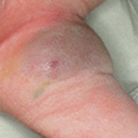
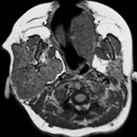
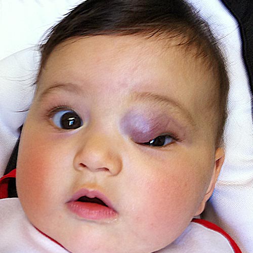
If the fast-flow properties of an arteriovenous malformation in childhood are not yet fully developed, a misdiagnosis of infantile hemangioma might be possible. Both an infantile hemangioma and an arteriovenous malformation are warm on palpation and reddish when there is skin involvement. However, an arteriovenous malformation in infancy appears as a faint capillary redness with teleangiectasia, while an infantile hemangioma has a strong strawberry color. Infantile hemangioma goes through classic phases, is not yet detectable at birth and proliferates, i.e., clearly increases in size, especially in the first year of life, whereas an arteriovenous malformation hardly expands in the first year of life and does not exhibit a tumor-like, solid appearance. This difference is also reflected on imaging: In contrast to an arteriovenous malformation, an infantile hemangioma appears on MRI as a focal, homogeneous, circumscribed solid mass with flow voids, especially in the proliferation phase.
In contrast to infantile hemangiomas, these hemangiomas are already fully developed and visible at birth. Postnatally, they either undergo rapid regression (RICH: rapidly involuting congenital hemangioma) or remain unchanged (NICH: non-involuting congenital hemangioma), but never increase in size. On account of the rapid regression in the first life year, an RICH can be readily distinguished clinically from an arteriovenous malformation.
The NICH is sometimes not immediately distinguishable from an arteriovenous malformation of small vessels. However, it appears as a clearly circumscribed solitary tumor. Another difference from an arteriovenous malformation is that an NICH grows proportionally with the growth of the child and usually appears as a voluminous dark red to bluish skin lesion surrounded by a pale halo. On MRI an NICH will look similar to an infantile hemangioma. Unlike an arteriovenous malformation, however, it demonstrates a tumor-like parenchymal blush. In addition, the afferent arteries are much smaller in caliber, and an immediate, early venous contrast outflow is not detectable.
In addition, a clinical distinction should be made between a simple arteriovenous malformation and an AVM-related syndrome with anomalies of other tissues or vessels (e.g., Parkes Weber syndrome, CM-AVM, CLOVES syndrome, HHT - Osler's disease).