Chapter: Invasive therapy
Article: 12 of 15
Update: Mar 08, 2021
Author(s): Kühnel, Thomas
The general principles of tumor surgery cannot be easily transferred to the surgery of malformations. Communication with the patient must be adapted to the very specific situation. Successful surgery of vascular malformations therefore requires the surgeon to rethink and adapt the procedures learned in oncological surgery.
At the latest during the informed consent procedure, the patient will be put through a process that is laborious and sometimes painful. The patient has to learn that the nature of his or her disease is often not entirely curable and that in most cases the goal is defined more modestly than the patient might have imagined. In addition, it may be necessary to discuss the fact that the treatment might require several sessions and that each intervention has clearly defined goals.. It is only by building up a realistic expectation in the patient that the usually very longstanding relationship between doctor and patient can be built on trust that is stable enough to survive disappointments.
In contrast to tumor surgery, where resection of the lesion within oncologically safe margins is the goal to which cosmetic and possibly functional aspects must be subordinated, other primary goals are defined in the surgical therapy of malformations.
Preservation or restoration of the best possible quality of life for as long a period as possible is the primary goal in the treatment of malformations. Such an objective is much more vaguely defined than in tumor surgery and should therefore always be explored and elaborated together with the patient. Especially in the head and neck region, the cosmetic aspect plays a major role and must be taken into account. It is even more important to weigh functional aspects wisely against radicality. It is sometimes wise to offer a young patient an incomplete resection of the lesion, knowing that follow-up procedures may have to be performed under less favorable conditions. The advantage of this strategy may be an external appearance that allows the patient to socialize better than if radical surgery were performed.
Another difference from tumor surgery can be found in the chronological order of the treatment steps. Whereas in oncology the first intervention usually determines the success of the therapy, surgery in partial steps is not only permissible but is necessary for a large number of malformations in order to achieve a good result. This requires a lot of understanding of the disease but also a lot of patience on the part of the patient.
The prerequisites for promising surgery of vascular malformations cover very different aspects and resources of medicine. Knowledge of hemostaseology is just as indispensable as profound experience in plastic and reconstructive surgery.
Beyond the expertise of the surgeon, intensive interdisciplinary exchange with a variety of specialist disciplines is essential. At the forefront is highly specialized interventional radiology which, in addition to diagnostics, is playing an increasingly important role in therapy. Modern pharmacological therapy with its immunologic implications should be performed in consultation with appropriately experienced colleagues in pediatrics, hematology and oncology. It is naturally important to ensure close cooperation with anesthesiologists and intensive care physicians, who not only take over postoperative care of potentially critically ill patients, but also have to be prepared intraoperatively for high blood loss. As well as the cell saver, different options for bleeding control must be provided, which are seldom used in other operations. Particularly in the case of arteriovenous malformations but also venous malformations, the surgical team must be aware that common methods for bleeding control such as ligation and electrocoagulation may fail. Compression and the local application of hemostyptic substances may be advisable. For the author, a cascade of measures that also takes the economic factor into account has proven helpful. For example, carboxymethyl cellulose (such as Tabotamp™), also in the form of microfibrils (Tabotamp Fibrillar™), is used primarily for local control of bleeding, while gelatin gels with thrombin such as Surgiflow™ or Floseal™ are used when the effect is inadequate.
For venous malformations with a risk of disseminated intravascular coagulation in large caverns, anticoagulation must be started well before the operation. In the case of large lesions, one or even more preoperative interventional therapies are usually extremely helpful in improving the overview of the surgical site and reducing the amount of bleeding. Multiple interventions are regularly indicated. Even more important in arteriovenous malformations, embolization is required to obtain a condition were surgery is possible. Sufficient embolization also helps to identify the limits of the arteriovenous malformation compared to normal tissue. For surgery, it is important to consider carefully which diagnostic imaging is available and whether blood products are available in sufficient quantities.
Even though approvals for anti-angiogenic drugs for the treatment of malformations are pending in Germany, their use has become increasingly established. There are still no consistent therapy recommendations but there is a broad consensus on the promising future of pharmacotherapy. This is also true for the treatment of lymphatic malformations, where surgery is an important, but by no means a central pillar of therapy.
Access to lesions in the face is based on the rules of plastic surgery. In addition to skin folds, the relaxed skin tension lines (RSTL) are guiding principles for access. If it is planned to remove the lesion with its epidermal parts completely, an incision outside the lesion is chosen. Thus unaffected skin is used during wound closure, promising undisturbed wound healing.
Flat lesions can also be removed sequentially. Starting in the center of the pathology, an ellipse or other area is resected that can be primarily closed. In the next step the procedure is repeated until the diseased area is completely resected. The high time expenditure and the necessity for repeated anesthesia is offset by the advantage of finally achieving wound closure with local tissue. It also makes it easier to check for recurrence.
In almost all cases, the wound surfaces are consistently treated with absorbent drains (Robinson). Even if the wound drainage rate drops rapidly postoperatively, drainage is typically provided for a period of four days. Inpatient observation may also be longer than in comparable soft tissue procedures without vascular malformation. We prefer moist wound healing and use foils for wound coverage. After removal of the foil and any pressure dressing that may be required, the patient treats the wound edges with moist ointments such as Vaseline. Sun blockers are recommended for at least six months to prevent discoloration of the skin.
Local flap surgery involves the issue of vascular supply at the flap margins with a risk of trophic disturbances, which is a serious problem in the context of difficult wound healing conditions. Therefore, flap techniques with axial vascularization should be preferred.
Preservation of the facial nerve is a primary goal for the functioning and esthetics of the face. The nerve is electrophysiologically monitored during surgery. Dissecting its peripheral parts in the vicinity of a malformation is technically extremely demanding and sometimes frustrating.
Penetration of the mimic musculature by pathological vascular tissue often leads to extensive resection of musculature, which means facialis function disappears anyway. This must be explained in advance. The same applies to secondary facial rehabilitation by static or dynamic procedures. Suspension of the corner of the mouth with temporalis fascia or Gore-Tex patch or the (orthodromic) temporal muscle transfer are available for this purpose. Very complex procedures such as the latter or even the microvascular gracilis muscle transfer must be discussed critically with the understanding that revision of the malformation might be necessary.
Intraoperative bleeding, especially in the case of arteriovenous malformations, is a situation for which the surgeon is prepared and should not be considered a complication but a normal situation. The classic hemostasis procedures such as electrocoagulation, transfusion and ligation are often not sufficient on their own and are supplemented by compression and adhesion of the wound surfaces. The situation is more difficult if a hematoma occurs postoperatively. In this case, it is necessary to decide when and whether to revise. Of course, even small hematomas can lead to infections and endanger the surgical result.
On the other hand, reopening the wound means further trauma at the wound margins, which will lead to a worse scar.
General surgical procedures are demonstrated using the example of some special situations:
In this case of an extensive venous malformation in the face, not only cosmetic disfigurement but also massive functional deficits occurred. These mainly concerned the intake of food. Imaging diagnostics confirmed the clinical examination findings, namely that the lesion had almost completely penetrated the mimic muscles of the left half of the face and that muscle function had failed in the area innervated by the facial nerve. To reduce the volume and the risk of bleeding, multiple sclerotherapies were performed preoperatively.
After drug therapy with sirolimus and interventional radiological therapy, surgical improvement of the situation was planned.
Different, sequential access routes were assessed. The pre-auricular part of the malformation was addressed via a parotid access with facial nerve monitoring. The mimic musculature was deeply involved.
In the next step, the perioral part was resected via an external access in the nasolabial fold and an intraoral access in the vestibulum oris.
The eyelid region and the nose are spared. The eyelid has sufficient function and would most likely show deficits after surgery. The same applies to the external nose. Its contour is largely determined by the volume of the malformation. The wing and lateral cartilages are very delicate or destroyed. There is a significant deviation that could only be corrected with rib cartilage augmentation. The soft tissue deficits would have to be covered with a frontal flap. All these measures are unnecessary in the current situation and would probably result in a serious deterioration in quality of life. Corrections to the corner of the mouth with static suspension complete the current treatment plan.
In this example an arteriovenous malformation is described, which was treated several times interventionally with EVOH and pharmacologically with sirolimus before surgical treatment. Complications such as profuse bleeding from the orbit, repeated bacterial infections as well as the disastrous social component made the patient decide to undergo surgery.
The orbit was completely infiltrated by the malformation, the eyelids were inoperable. Resection and reconstruction while preserving the eye proved to be impossible. Therefore, the orbital portion of the AVM was completely embolized preoperatively including the eye.
Removal of the complete orbital content and defect coverage with microvascular pedicled latissimus dorsi flap were planned.
The part of the malformation that crossed the midline was left to a second procedure to preserve the contour of the outer nose. Subsequently, the flap was thinned out to such an extent that an epithesis was possible.
Postoperatively, not only did the patient’s general condition improve because there was no more bleeding or infection. The psychological condition also showed a profound improvement.
This example shows that, after an operation 15 years earlier, revision is possible with equal success. The reappeared arteriovenous malformation, which had gained in dynamics in the time before the operation demonstrated here, was covered with a local flap plasty after embolization. To cover the expected tissue deficit without tension, a skin expander was inserted close to the malformation and successively filled. The diseased tissue was removed and covered with a local flap, taking into account the position of the eyebrows.
Postoperatively, the patient experienced a good cosmetic and functional result.
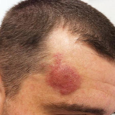
The protruding and pulsating ear was an unbearable condition for the patient. The increase in size did not begin until adolescence. The angiography demonstrates the large-caliber feeding vessel, which is also striking during surgery. The nidus was resected in toto, and the wound was primarily closed. An anterior approach was used to reduce the size of the helix in the same session.
Lymphatic malformations can lead to functional loss if the tongue is affected, as in the example shown. The increase in volume prevents the patient from closing the mouth completely, resulting in continued sialorrhea. Mastication as well as articulation is impaired. If drug therapy (e.g., with sirolimus systemically) or sclerotherapy (e.g., with Bleomycin locally), does not produce the desired success, wedge resection may improve the situation for a long period of time. The tissue resections laterally at the tip of the resectate in the body of the tongue allow the approximation of the tissue margins without bulging. The disadvantage of this method is a loss of sensitivity in the scarred area. In this situation, however, the motor function as well as the sensitivity of the tongue is usually seriously reduced in any case.
At first glance not a dramatic situation, this microcystic lymphatic malformation of the right half of this boy’s face was nevertheless seen as an indication for surgery. Not only did it represent a cosmetic impairment, it also led to altered bone growth of the jaw with corresponding functional deficits.
After unsuccessful drug therapy, an extensive resection was attempted.
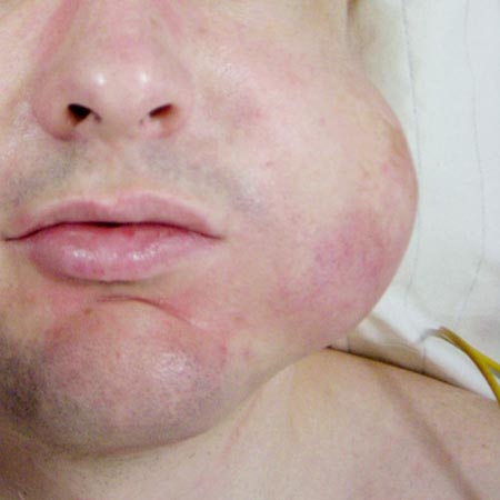
Perioral capillary malformations may be approached for cosmetic reasons, as they can be accompanied by soft tissue hyperplasia with increasing age. Here, too, it is preferable to proceed step by step rather than to induce incalculable scars or destroy the function of the orbicularis oris muscle. Even if it is possible to improve the profile of the perioral region, color matching will not succeed surgically. Vermilion plasty is not an option because the vestibular mucosa has the same coloration as the lips. Laser therapy is the method of choice here.
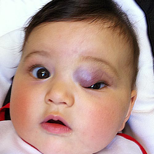
Malformations of the periorbital region are probably the most difficult surgical challenge. The delicate structures of the lower eyelid, in particular, are hardly forgiving of scar contractures; any tissue resection requires difficult, careful reconstruction. We operate on malformations of the eyelids only in trusting cooperation with the ophthalmologist performing the plastic surgery. A combined approach is advantageous because venous, or even more so arteriovenous malformations do not respect the structural elements of the eyelids such as the tarsus and the retractors of the lower eyelid. We start with the transconjunctival approach and operate from the outside. It is important to avoid a malposition of the lacrimal point in order not to cause epiphora. If the tarsus has to be resected or is destroyed by the pathology, reconstruction can be performed in the same session with septal or auricular cartilage. Whenever possible, defects of the epidermis are avoided. If free skin grafts are required, they should be taken from a region of similar color and texture. Retroauricular skin is a suitable option. The local flap of eyelid surgery is used for defects at the edge of the eyelid. In this regard, reference is made to the extensive ophthalmoplasty literature.